
INTRODUCTION
Bladder cancer was the sixth most commonly diagnosed cancer in males in 2012 worldwide and the most common malignancy of the urogenital tract. In 2012, 429,800 new cases of bladder cancer were reported worldwide, meanwhile 165,100 patients died from the disease [1].
Protein kinase casein kinase 2 (CK2) is a conserved, ubiquitously expressed protein serine/threonine kinase that was first discovered by Burnett and Kennedy in 1954 [2]. CK2 exists primarily as a holoenzyme consisting of two catalytic subunits, CK2α and CK2α’, and two β regulatory subunits. Functionally, CK2 is involved in the modulation of major cellular processes and pathways [3, 4]. Subsequent studies have shown that CK2 was a potent regulator in the processes of cell proliferation, differentiation, and apoptosis, which was involved in numerous signaling pathways including PI3K/AKT, NF-κB, Wnt, Notch1 and Hedgehog/Gli1 [5–9]. Although its pro-survival function is necessary for the whole organism, it is ‘more necessary’ for cancer cells than normal. Elevated level of CK2α has been observed in numerous cancers, including breast [10], prostate [11], lung [12, 13], head and neck [14], colorectal [15], gastric [16], and kidney [17]. These evidences validated that CK2 could be a potential cancer therapeutic target. CX-4945 was the first effective and selective CK2 inhibitor used in human clinical trials. CX-4945 inhibits the activity of CK2α and CK2α’, resulted to suppression of activation of pro-survival signaling pathways and promotion of apoptosis in human glioblastoma, breast, prostate, and lung cancers [13, 18–20].
Normal cells metabolize glucose to produce ATP by oxidative phosphorylation and a large amount of pyruvate which is oxidized via the tricarboxylic acid cycle. The rate of production is coupled with oxygen respiration and proton transport [21]. By contrast, tumor cells consume glucose to produce lactate and support mitochondrial oxidative phosphorylation even in oxygen-rich conditions. This phenomenon is referred to as “aerobic glycolysis” or the “Warburg effect” which was first observed in the 1930s by Otto Warburg [22]. Increased glycolysis is widely considered as a crucial hallmark of cancer, which has been consistently observed in many cancer cell lines [23]. Recent studies indicated that cellular metabolism was directly or indirectly linked to activated oncogenes and inactivated cancer suppressors [23, 24]. The expression level of glycolysis-related genes, such as glucose transporter (GLUT)1, lactic dehydrogenase (LDH)A, LDHB, hexokinase (HK)1, HK2, and pyruvate kinase type M (PKM), are elevated in certain cancer tissues [25], which have been observed in our previous work. Thus, enhanced dependency of cancer cells on glycolysis probably provides a biochemical basis to kill malignant cells preferentially than normal cells by inhibiting glycolysis.
Above all, we reported the function of CK2α in bladder cancer and further investigated the possible relationship between CK2α and glucolysis. We also tested the potential anticancer effect of inhibition of CK2 in vitro and vivo, suggesting its possible application in the treatment of bladder tumors.
RESULTS
Overexpression of CK2α in human bladder cancer
Primary paired BCa tissue samples and BCa cell lines were used to examine CK2α expression. The gene expression of CK2α was analyzed by qRT-PCR in 22 pairs of BCa tissues and their adjacent tissues. Compared with adjacent bladder tissues, the mRNA level of CK2α was significantly overexpressed in the bladder tumor tissues (P < 0.01; Figure 1A and 1B). To investigate whether differences in expression of mRNA level are reflected at the protein level, Western blot analysis was conducted. Consistent with the PCR results, CK2α protein expression was significantly higher in the BCa tissues compared with that in the adjacent tissues (Figure 1C). qRT-PCR analysis demonstrated that mRNA level of CK2α in six bladder cancer cell lines (T24, J82, EJ, 253J, TCC, and RT4) was increased (particularly in T24) compared with that in the normal urinary epithelial cell line sv-Huc (Figure 1D). Thus, CK2α expression was positively associated with bladder cancer progression, suggesting that it played an oncogenic role in bladder cancer.
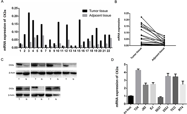
Figure 1: Expression level of CK2α mRNA and protein in human primary bladder cancer cell lines and surgical specimens as evaluated. A, B. RT-qPCR showed that relative CK2α mRNA expression in 22 BCa and adjacent tissues (P < 0.05). C. CK2α protein in seven BCa and adjacent tissues (N, non-tumor, T, tumor). D. CK2α mRNA levels was up-regulated in seven bladder cancer cell lines compared with those in the normal urinary epithelial cell line sv-Huc.
IHC analysis of CK2α expression in BCa clinical samples
To further explore the role and prognostic value of CK2α in human bladder cancer, TMAs with 160 cases of bladder tumor samples were used to examine CK2α expression via IHC. In the CK2α-positive specimens, high CK2α expression (++ or +++) was found in 101 (63.1%) specimens, and low CK2α expression (− or +) was detected in 59 (36.9%) specimens (Table 1). Correlations between the clinicopathological parameters of BCa and expression of CK2α are summarized in Table 1. Chi-square analyses revealed that CK2α expression was positively correlated with histological grade (P = 0.024) but not with tumor size and tumor stage (TNM). Kaplan–Meier analyses did not reveal a significant association between high CK2α expression and poor prognosis(P = 0.694; Figure 2B).
Table 1: Correlation between CK2α expression and clinicopathological variables of 160 patients with bladder cancer.
Clinicopathologic variable |
CK2α expression |
|||
|---|---|---|---|---|
N |
Low |
High |
P |
|
All cases |
160 |
59 |
101 |
|
Age(years) |
0.373 |
|||
<65 |
74 |
30 |
44 |
|
≥65 |
86 |
29 |
57 |
|
Gender |
0.970 |
|||
Male |
125 |
46 |
79 |
|
Female |
35 |
13 |
22 |
|
TNM stage |
0.308 |
|||
Stage I |
92 |
37 |
55 |
|
Stage II and III |
68 |
22 |
46 |
|
Histological grade |
||||
grade I and II |
79 |
36 |
43 |
0.024* |
grade III |
81 |
23 |
58 |
|
Tumor size(cm) |
0.611 |
|||
<3 |
110 |
42 |
68 |
|
≥3 |
50 |
17 |
33 |
|
*P < 0.05
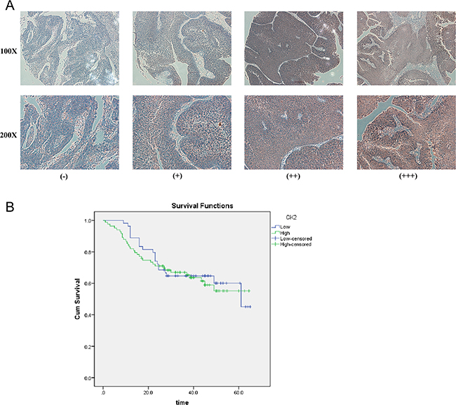
Figure 2: IHC analyses of CK2α protein expression in primary BCa surgical specimens and Kaplan–Meier survival analyses of the primary BCa patients (n = 137). A. CK2α staining in BCa, scored as CK2α (-) (+) (++) (+++). All images are shown at × 100 magnification and × 200 magnification. B. BCa patients were divided into low-CK2α expression (n = 54, CK2α- or CK2α+) and high-CK2α expression (n = 83, CK2α++ or CK2α+++) groups. Survival analyses did not reveal a significant association between high CK2α expression and poor prognosis (P = 0.694, log-rank test).
CK2α promotes bladder cancer cell proliferation
Cell proliferation assay was conducted to explore the role of CK2α in the growth of bladder cancer cells. T24 and EJ cells were evaluated in cell proliferation assays after they were transiently transfected with CK2α-specific siRNAs and siNC RNA (negative control, NC) for 48 h,. Cell proliferation (P < 0.05; Figures 3D and 3E) was significantly inhibited in T24 and EJ cells transiently transfected with siCK2α compared with those transfected with siNC. These results supported that CK2α promoted the growth of BCa cells.
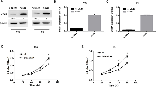
Figure 3: CK2α was essential for bladder cancer cell proliferation. A, B, C. Knockdown efficiency of selected CK2α-targeting siRNAs in transfected cells was evaluated by Western blot and qRT-PCR. D, E. CCK8 assay showed that silencing of CK2α suppressed proliferation of T24 (D) and EJ (E) cell lines.
Inhibition of CK2α downregulates the glucose metabolic pathway
To investigate whether CK2 suppression influences the glucose metabolic pathway, we silenced CK2α expression. At 48 h after transfection, the efficiency of RNA interference was monitored by Western blot and qRT-PCR. The corresponding expression of CK2α in T24 and EJ cell lines was decreased (Figures 3A, 3B, and 3C). Glucose uptake and lactate secretion in bladder cancer cells decreased when CK2α was inhibited (Figures 4A and 4B). AKT was found to be less phosphorylated at Ser473 in CK2α-silenced cells than in control cells (Figure 4C). AKT is considered the Warburg kinase. PI3K/AKT/mTOR signaling links several tumor cell metabolic processes, including glycolysis. As such, we found that the protein level of glycolysis-related genes, including GLUT1, G6PD, LDHA, LDHB, HK1, HK2, and PKM, was lower in CK2α-silenced cells compared with those in control cells (Figure 4C). Furthermore, treatment with CX-4945 suppressed glucose uptake and lactate secretion (Figures 5F and 5G) in bladder cancer cells. AKT was less phosphorylated at Ser473, and the protein level of glycolysis-related genes was lower in cells after treatment with CX4945 (Figures 5C and 5E). Taken together, CK2α inhibition may suppress high glycolysis levels via suppressing AKT phosphorylation in bladder cancer cells.
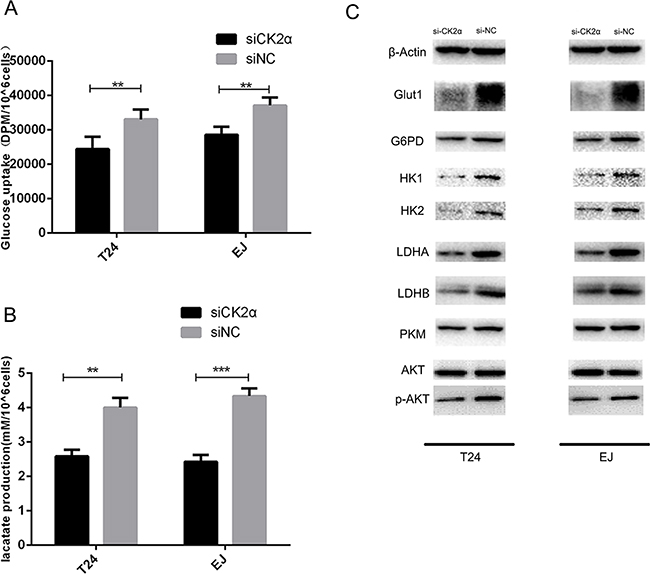
Figure 4: CK2α inhibition induced a metabolic shift in bladder cancer cells. A. Glucose uptake assays. CK2α inhibition decreased glucose uptake. B. Lactate production assays. CK2α inhibition suppressed lactate secretion. C. CK2α inhibition decreased protein expression in cancer aerobic glycolysis.
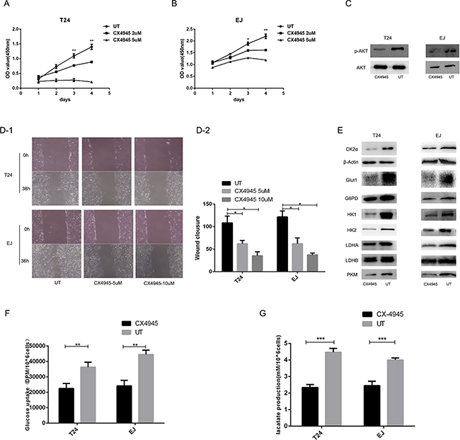
Figure 5: Effects of inhibition of CK2 on bladder cancer cell function. A, B. CX-4945 suppressed proliferation of T24 (A) and EJ (B) cell. C, E. Western blot analysis showed that protein expression in cancer aerobic glycolysis was downregulated in T24 and EJ cells after treatment with CX4945. AKT and p-AKT(ser473) (C). GLUT1, G6PD, HK1, HK2, LDHA, LDHB and PKM (E). D. CX-4945 inhibited the migration ability in T24 and EJ cell lines. F. Glucose uptake assays. CX-4945 decreased glucose uptake. G. Lactate production assays. CX-4945 suppressed lactate secretion.
Effects of inhibition of CK2 on BCa cell function
Treatment with CX-4945 suppressed bladder cancer cell growth in a dose-dependent manner (Figures 5A and 5B). We found that treatment with CX-4945 significantly inhibited the migration ability in bladder cancer cells (Figure 5D; both P < 0.05). CX-4945 induced apoptosis in both cell lines (Figure 6; both P < 0.05). Thus, CK2 inhibition abrogated many biological functions of bladder cancer that are critical to tumor growth and survival.
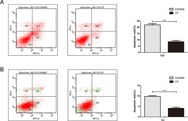
Figure 6: Effect of inhibition of CK2 on apoptosis in bladder cancer cells. CX-4945 induced apoptosis in T24(A) and EJ(B) cell lines.
Knockdown of CK2α inhibits the growth of tumor xenografts in nude mice
We evaluated the effects of CK2α Knockdown on tumor growth in vivo. The transfected EJ cells were implanted into the right axilla of the nude mouse by subcutaneous injection, and the tumor volumes were measured after injection. Compared with the control cells, the mean volumes of the xenograft tumors treated with CK2α knockdown cells showed obvious growth retardation (Figure 7A, 7B, 7C, 7D). Immunohistochemical staining identified that the level of CK2α was inhibited in the CK2α knockdown tumors (Figure 7E).
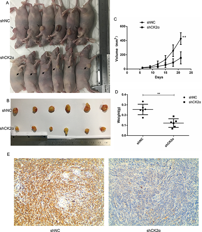
Figure 7: CK2α inhibition decreased tumorigenesis and reduced cancer cell growth in vivo. A, B. CK2αknockdown EJ cells formed smaller tumour volume compared to the control cells (NC). C. The tumor growth curves in the two groups (P < 0.01). D. CK2αknockdown reduced tumor weight compared to the control cells (NC) (P < 0.01). E. CK2α immunohistochemistry staining of xenografted tumors retrieved from treated nude mice. Representative images are shown. (magnification, ×100)
DISCUSSION
Protein kinase CK2α, one isoform of the catalytic subunit of serine/threonine protein kinase CK2, has been demonstrated to be overexpressed in various malignancies [26–28]. However, its underlying role in bladder cancer remains unclear.
In this study, we revealed that CK2α was frequently and significantly upregulated in human bladder cancer. The level of CK2α in bladder cancer cell lines except 5637 was increased compared with that in the normal urinary epithelial cell line sv-Huc. The difference results might be caused by low malignance of 5637 or heterogeneity of tumor. We expect further research on this issue. IHC analyses convincingly demonstrated that CK2α was overexpressed in primary bladder cancer. Increased expression of CK2α was shown in 63.1% of BCa samples, and was significantly correlated with tumor grade. These findings indicated that CK2α was an oncogene promoting bladder cancer progression and correlated with pathogenesis.
As a prognostic marker in various malignancies, CK2α expression is positively correlated with histological grade, distant metastasis, tumor stage and outcome [15, 27, 29]. However, we found no significant evidence to identify the relationship between high CK2α expression and tumor stage and prognosis. These differences might be attributed to tumor heterogeneity.
Numerous signaling pathways have been researched, through which CK2 regulates cancer cell survival, but the relationship between CK2 and glucose metabolism remains unknown. Our present work suggested that CK2 may be a positive regulator in cell survival via the glucose metabolic pathway in bladder cancer. Several lines of evidence support this finding. Firstly, the inhibition of CK2α resulted in the downregulation of glycolysis-related protein level, as well as decrease in glucose uptake and lactate production. Secondly, suppressed cell proliferation was observed after CK2α knockdown. Finally, similar results were reproduced after treatment with CX-4945 even with a dose-dependent response in both cell lines.
Our previous study demonstrated that the expression level of the glycolysis-related genes in bladder tumor tissues was frequently overexpressed compared with those in adjacent tissues [25]. Through the glucose metabolic pathway, tumor cells consume glucose to produce a large number of ATP, lactate, NAD+, NADPH, and H+ cofactors, which are used for the growth and proliferation of cancer cells. In our study, we knocked down CK2α level in bladder cancer cells and then observed decrease in glucose uptake and lactate production, accompanied with low level of GLUT1, HK, PKM, and LDH. These results suggested that CK2α positively regulated the activation of glycolysis. Interestingly, CK2α silencing reduced the expression level of G6PD, which was of great importance for the pentose phosphate pathway (PPP). PPP is a key source of NADPH, as a cofactor in maintaining the redox balance and for anabolic pathways, such as fatty acid biosynthesis [30]. Moreover, PPP produces ribose and is involved in the transcription of gene expression during stress conditions [31].
In human glioblastoma, CK2 suppresses apoptosis and promotes oncogenes via enhancing the AKT pathways and downstream gene expression [18]. Consistent with these observations, AKT was found to be less phosphorylated in CK2α knockdown cells than in control cells, which suggested that CK2α-regulated glycolysis was related to the AKT pathway. Several reports conveyed the ability of CK2 inhibitors to suppress the PI3K-AKT-mTOR pathway [32–34]. Dr. Ruzzenne demonstrated that CK2 phosphorylates AKT directly at Ser129, further increasing the catalytic activity of AKT [35]. Unsurprisingly, hyper activation of PI3K-AKT-mTOR signaling was shown to be a consequence of CK2 overexpression [36, 37]. Researchers have found that the PI3K-AKT-mTOR axis was an essential pathway for various cellular processes, including glycolysis [38, 39]. Moreover, AKT has been reported to function as a master regulator of energy metabolism in cancer cells by enhancing the expression of glycolytic regulators [38]. Taken together, CK2α inhibition may suppress high glycolysis level via the CK2α/AKT-mTOR cascade in bladder cancer cells.
Notably, bladder cancer cell treatment with CX-4945 strongly inhibited activation of AKT phosphorylation and glycometabolism. After treatment with CX-4945, it were observed that suppressed cell proliferation and cell migration and induced cell apoptosis. Thus, CK2 inhibitors, such as CX-4945, may provide a potential therapeutic target for patients with bladder cancer. Moreover, in vivo xenograft study showed that knockdown of CK2α decreased the tumorigenicity of bladder cancer cells.
MATERIALS AND METHODS
Clinical specimens and cell culture
Bladder cancer and adjacent non-tumor tissues were obtained from patients undergoing radical cystectomy at the First Affiliated Hospital of Nanjing Medical University. All patients provided signed informed consent, and the Research Ethics Committee of our institution approved this study. The specimens were immediately frozen and stored in liquid nitrogen. Pathological examination was performed to confirm cancer diagnosis.
Bladder cancer cell lines T24 and EJ were obtained from the Type Culture Collection of the Chinese Academy of Sciences (Shanghai, China). Cells were cultured in RPMI 1640 medium supplemented with 10% fetal bovine serum (FBS; Gibco, Australia) and 1% penicillin–streptomycin in an incubator with humidified 5% CO2 at 37°C.
All the bladder cancer specimens we used in our work are urothelial carcinomas. The cell type of T24, J82, EJ, 5637, 253J and TCC is Transitional Cell Carcinoma, while the cell type of RT4 is Transitional Cell Papilloma.
Tissue microarray (TMA) and Immunohistochemistry (IHC)
TMA (16×10) was constructed from 160 cases of bladder tumor tissues. TMAs were kept at 4°C until they were ready for analysis.
Sections were arranged in duplicate cores per case. TMAs were treated with xylene and 100% ethanol, followed by decreased concentrations of ethanol. After antigen retrieval, TMAs were blocked and stained with antibodies against CK2α, followed by secondary antibody incubation and standard avidin biotinylated peroxidase complex method. Hematoxylin was used for counterstaining, and images were obtained with an upright microscope system (Nikon, JAPAN).
The total CK2α immunostaining score was calculated as the sum of the score for the proportion of positively stained tumor cells and the score for staining intensity given by two pathologists blinded to the clinical parameters. The proportion of positively stained tumor cells was scored as follows: “0” (<5%, negative), “1” (5%–25%, sporadic), “2” (25%–50%, focal), and “3” (>50%, diffuse). The intensity of staining was graded according to the following criteria: “0” (no staining), “1” (weak staining = light yellow), “2” (moderate staining = yellow brown), and “3” (strong staining = brown). The total immunostaining score, which ranged from 0 to 9, was calculated as the value of the proportion of positive cell score × staining intensity score. The expression level of CK2α was defined as follows: “−” (negative, score 0), “+” (weakly positive, score 1–3), “++” (positive, score 4–6), or “+++” (strong positive, score 7–9). Thus, CK2α protein expression in BCa specimens was divided into two groups: low CK2α expression group (CK2α“–” or CK2α“+”) and high CK2α expression group (CK2α“++” or CK2α“+++”) (Figure 2A).
CK2α inhibitor
CK2α siRNA and control siRNA were purchased from Shanghai GenePharma Co., Ltd. Cells were seeded in a six-well plate at 10^5 cells/well 1 day before transfection, with a target of 30%–50% confluence at the time of transfection. Cells were transfected with 50 nmol/L siRNA using Lipofectamine RNAiMAX (Invitrogen) according to the manufacturer’s protocol. Adequate inhibition of siRNA-mediated knockdown was confirmed by Western blot. The pcDNA3.1- CK2α or control pcDNA3.1-LacZ plasmid vectors were then transfected into T24 and EJ cells (0.5l g/mL in a 24-well plate) using Lipofectamine 2000 transfection reagent (Invitrogen), according to the manufacturer’s protocol. Cells were harvested for qRT-PCR and Western blot or used in other assays at 48 h post-transfection.
Lentivirus constructs were generated to knockdown CK2α. The bladder cancer cell EJ was stably transfected with LV3pGLV-h1-GFP-puro negative control vectors (termed as shNC) and CK2α knockdown lentivirus (termed as shCK2α), following the manufacturer’s instructions (Abm, Nanjing, China). Lentiviral constructs of CK2α knockdown were generated as previously described. Cells were plated in 6 wells dishes at 40% confluence and infected with the retroviruses. Meanwhile, polybrene (3 μg/ml) was added with the retroviruses to enhance infection efficiency. Stable pooled populations of bladder cancer cells were generated by selection using puromycin (3μg/ml) for 3 weeks. For CK2α knockdown cells, which achieved≥75% knockdown efficiency of mRNA was used for further studies.
CX-4945(small-molecule CK2 inhibitor) was purchased from Selleckchem.
RNA extraction and quantitative RT-PCR
Total RNA from 44 frozen bladder tissues (22 for tumor tissues and 22 for paired adjacent normal tissues) and bladder cancer cells was extracted by Trizol according to the manufacturer’s instructions. cDNA was prepared from 1 μg of total RNA in a 30 μL volume using PrimeScriptTM RT Master Mix (Takara Bio, Dalian, China). The reaction mixture was incubated at 37°C for 15 min, 85°C for 5 s, and 4°C for 5 min. The synthesized cDNA was used for PCR amplification or stored at −20°C for further analysis.
The PCR reaction was performed in a 25 μL volume containing 0.25 mM of each dNTP, 1 U HotstarTaq (Takara), and 0.5 mM of each primer (Takara): 5′-CCGAGTTGCTTCCCGATAC-3′ (forward) and 5′-GGGCTGACAAGGTGCTGAT-3′ (reverse) for CK2α; 5′-AGCGAGCATCCCCCAAAGTT-3′ (forward) and 5′-GGGCACGAAGGCTCATCATT′ (reverse) for β-actin.
Western blot analysis
Cells were lysed, and the protein was extracted using RIPA buffer (Beyotime, China) and quantified by a BCA Protein Assay Kit (Beyotime, China). Equivalent quantities of protein were separated on 10% SDS–PAGE gels and transferred to polyvinylidene fluoride membranes. After blocking with 5% nonfat milk at room temperature for 1 h, the membranes were immunostained with primary antibodies at 4°C overnight, washed three times in TBST, and incubated with secondary antibody at room temperature for 1 h. Band signals were detected using a chemiluminescence system (Bio-Rad, USA) and analyzed using Image Lab Software. The following primary antibodies were used: primary antibodies against GLUT1, LDHA, LDHB, G6PD, 6PGD, and PKM2 were obtained from Epitomics, whereas those against HK1, HK2, AKT, and phospho-AKT(Ser473) were obtained from Cell Signaling Technology. CK2α was obtained from Millipore. The protein levels were normalized to β-actin (1:1000, Cell Signaling Technology).
Measurement of glucose uptake
For glucose uptake experiments, cells were plated in six-well plates at a density of 3×10⁵ cells/well at 24 h before the experiment was performed. Cells were then washed with PBS, and uptake was initiated by incubating cells in 2 mL of Krebs–Ringer–HEPES (KRH) buffer (25 mMHepes, pH 7.4, 120 mMNaCl, 5 mMKCl, 1.2 mM MgSO4, 1.3 mM CaCl2, and 1.3 mM KH2PO4) containing 1 μCi of [3H]-2-deoxyglucose (PerkinElmer Life Sciences) for 20 min. Uptake was stopped by washing the cells with ice-cold KRH buffer. Cells were dissolved in 300 μL of lysis buffer (10 mMTris–HCl, pH 8.0, 0.2% SDS). Liquid scintillation spectrometry was performed to determine the radioactivity level of each aliquot. Disintegrations per minute value was used to evaluate the intracellular level of [3H]-2-deoxyglucose, and each assay was performed in triplicate.
Measurement of extracellular lactate
Approximately 3×10⁵ cells/well were seeded into six-well plates that were incubated in 2 mL of RPMI 1640 with 10% FBS. After incubation for 24 h, the supernatant was collected. The lactate concentration in the supernatant was quantified by performing a fluorescence-based assay according to the manufacturer’s instructions (L-Lactate Assay Kit, Cayman). The lactate concentration in the supernatant without seeded cells was also calculated and used to eliminate the effect of lactate on RPMI 1640 medium. Each assay was performed in triplicate.
Cell viability assay
For cell viability assay, the transfected cells were seeded into 96-well plates at a density of 3000 cells/well. The CCK8 method was used to determine cell viability at 24, 48, 72, and 96 h after the cells were seeded. The absorbance was measured at 450 nm using a Tecan Infinite F200 microplate reader.
Wound healing assay
Cells were seeded into six-well plates and cultured until they reached 95% confluence. A 200 μL pipette tip was used to generate a cross-shaped wound through the center of the well. The cultures were washed with PBS to remove cell debris, and the cells were incubated in RPMI 1640 medium without FBS. The wound was observed under a microscope (Olympus, Japan) at 40× magnification at two preselected time points (0 and 36 h), and the widths of wounds were counted.
Apoptosis assay
After treatment with CX-4945 for 12 h, cells were trypsinized, stained with Annexin V and propidium iodide, and examined by flow cytometry.
In vivo tumor xenograft studies
BALB/C female nude mice (4-6-weeks old, 18–22 g) were randomly divided into two groups (each containing 6 mice). The transfected EJ cells and controls were implanted with 1 × 10⁷ cells in 0.1 ml PBS per site on the axilla in nude mice (n=6). Tumor growth was monitored by measuring the width (W) and length (L) with calipers every 3 days, and the volume (V) of the tumor was calculated using the formula V=(W2×L)/2. After 21 days, mice were sacrificed and checked for final tumor size. At the end of the experiment, the tumors were removed and fixed in 4 % formalin for immunohistochemical analysis. The animal studies were performed in accordance with the institutional ethics guidelines for animal experiments.
Statistical analysis
Statistical analysis was performed using SPSS 20.0 and GraphPad Prism 5. All data are presented as the mean ± standard deviation. Differences between two groups were analyzed using Student’s t-test. Cell viability assay and in vivo tumor xenograft results were assessed by repeated measures ANOVA. P < 0.05 was considered statistically significant. All experiments were repeated more than three times, and each experiment was performed in triplicate.
CONCLUSIONS
Our study revealed the relationship between CK2α overexpression and unfavorable progression in bladder cancer patients. For the first time, we reported that targeting CK2 suppressed bladder cancer cell survival via downregulation of AKT-mTOR and the glucose metabolic pathway in urinary bladder cancer. However, the precise mechanism needs to be elucidated. Our results could be regarded as the basis for a candidate prognostic biomarker and new potential therapeutic strategies for bladder cancer. Our study also provided new insights into the regulation of glycolysis by protein kinase CK2.
ACKNOWLEDGMENTS AND FUNDING
This work was supported by grants from the National Natural Science Foundation of China (Grant Nos. 81272832 and 81602235) and Innovation Project for Postgraduate of Jiangsu Province (KYLX16-1110).
CONFLICTS OF INTEREST
The authors declare that no competing interests exist.
REFERENCES
1. Torre LA, Bray F, Siegel RL, Ferlay J, Lortet-Tieulent J and Jemal A. Global cancer statistics, 2012. Cancer J Clin. 2015; 65:87-108.
2. Burnett G and Kennedy EP. The enzymatic phosphorylation of proteins. The Journal of biological chemistry. 1954; 211:969-980.
3. Litchfield DW. Protein kinase CK2: structure, regulation and role in cellular decisions of life and death. Biochem J. 2003; 369:1-15.
4. Trembley JH, Wang G, Unger G, Slaton J and Ahmed K. Protein kinase CK2 in health and disease: CK2: a key player in cancer biology. Cell Mol Life Sci. 2009; 66:1858-1867.
5. Duncan JS and Litchfield DW. Too much of a good thing: the role of protein kinase CK2 in tumorigenesis and prospects for therapeutic inhibition of CK2. Biochim Biophys Acta. 2008; 1784:33-47.
6. Dominguez I, Sonenshein GE and Seldin DC. Protein kinase CK2 in health and disease: CK2 and its role in Wnt and NF-kappaB signaling: linking development and cancer. Cell Mol Life Sci. 2009; 66:1850-1857.
7. Guerra B. Protein kinase CK2 subunits are positive regulators of AKT kinase. Int J Oncol. 2006; 28:685-693.
8. Zhang S, Long H, Yang YL, Wang Y, Hsieh D, Li W, Au A, Stoppler HJ, Xu Z, Jablons DM and You L. Inhibition of CK2alpha down-regulates Notch1 signalling in lung cancer cells. J Cell Mol Med. 2013; 17:854-862.
9. Zhang S, Yang YL, Wang Y, You B, Dai Y, Chan G, Hsieh D, Kim IJ, Fang LT, Au A, Stoppler HJ, Xu Z, Jablons DM and You L. CK2alpha, over-expressed in human malignant pleural mesothelioma, regulates the Hedgehog signaling pathway in mesothelioma cells. J Exp Clin Cancer Res. 2014; 33:93.
10. Romieu-Mourez R, Landesman-Bollag E, Seldin DC, Traish AM, Mercurio F and Sonenshein GE. Roles of IKK kinases and protein kinase CK2 in activation of nuclear factor-kappaB in breast cancer. Cancer research. 2001; 61:3810-3818.
11. Laramas M, Pasquier D, Filhol O, Ringeisen F, Descotes JL and Cochet C. Nuclear localization of protein kinase CK2 catalytic subunit (CK2alpha) is associated with poor prognostic factors in human prostate cancer. Eur J Cancer. 2007; 43:928-934.
12. Daya-Makin M, Sanghera JS, Mogentale TL, Lipp M, Parchomchuk J, Hogg JC and Pelech SL. Activation of a tumor-associated protein kinase (p40TAK) and casein kinase 2 in human squamous cell carcinomas and adenocarcinomas of the lung. Cancer research. 1994; 54:2262-2268.
13. So KS, Rho JK, Choi YJ, Kim SY, Choi CM, Chun YJ and Lee JC. AKT/mTOR down-regulation by CX-4945, a CK2 inhibitor, promotes apoptosis in chemorefractory non-small cell lung cancer cells. Anticancer Res. 2015; 35:1537-1542.
14. Faust RA, Gapany M, Tristani P, Davis A, Adams GL and Ahmed K. Elevated protein kinase CK2 activity in chromatin of head and neck tumors: association with malignant transformation. Cancer Lett. 1996; 101:31-35.
15. Lin KY, Tai C, Hsu JC, Li CF, Fang CL, Lai HC, Hseu YC, Lin YF and Uen YH. Overexpression of nuclear protein kinase CK2 alpha catalytic subunit (CK2alpha) as a poor prognosticator in human colorectal cancer. PLoS One. 2011; 6:e17193.
16. Lin KY, Fang CL, Chen Y, Li CF, Chen SH, Kuo CY, Tai C and Uen YH. Overexpression of nuclear protein kinase CK2 Beta subunit and prognosis in human gastric carcinoma. Ann Surg Oncol. 2010; 17:1695-1702.
17. Stalter G, Siemer S, Becht E, Ziegler M, Remberger K and Issinger OG. Asymmetric expression of protein kinase CK2 subunits in human kidney tumors. Biochem Biophys Res Commun. 1994; 202:141-147.
18. Zheng Y, McFarland BC, Drygin D, Yu H, Bellis SL, Kim H, Bredel M and Benveniste EN. Targeting protein kinase CK2 suppresses prosurvival signaling pathways and growth of glioblastoma. Clinical cancer research. 2013; 19:6484-6494.
19. Siddiqui-Jain A, Drygin D, Streiner N, Chua P, Pierre F, O’Brien SE, Bliesath J, Omori M, Huser N, Ho C, Proffitt C, Schwaebe MK, Ryckman DM, Rice WG and Anderes K. CX-4945, an orally bioavailable selective inhibitor of protein kinase CK2, inhibits prosurvival and angiogenic signaling and exhibits antitumor efficacy. Cancer research. 2010; 70:10288-10298.
20. Pierre F, Chua PC, O’Brien SE, Siddiqui-Jain A, Bourbon P, Haddach M, Michaux J, Nagasawa J, Schwaebe MK, Stefan E, Vialettes A, Whitten JP, Chen TK, Darjania L, Stansfield R, Bliesath J, et al. Pre-clinical characterization of CX-4945, a potent and selective small molecule inhibitor of CK2 for the treatment of cancer. Mol Cell Biochem. 2011; 356:37-43.
21. Hinkle PC. P/O ratios of mitochondrial oxidative phosphorylation. Biochim Biophys Acta. 2005; 1706:1-11.
22. Warburg O. On the origin of cancer cells. Science. 1956; 123:309-314.
23. Cairns RA, Harris IS and Mak TW. Regulation of cancer cell metabolism. Nat Rev Cancer. 2011; 11:85-95.
24. Kroemer G and Pouyssegur J. Tumor cell metabolism: cancer’s Achilles’ heel. Cancer Cell. 2008; 13:472-482.
25. Yang X, Cheng Y, Li P, Tao J, Deng X, Zhang X, Gu M, Lu Q and Yin C. A lentiviral sponge for miRNA-21 diminishes aerobic glycolysis in bladder cancer T24 cells via the PTEN/PI3K/AKT/mTOR axis. Tumour Biol. 2015; 36:383-391.
26. Zhang S, Wang Y, Mao JH, Hsieh D, Kim IJ, Hu LM, Xu Z, Long H, Jablons DM and You L. Inhibition of CK2alpha down-regulates Hedgehog/Gli signaling leading to a reduction of a stem-like side population in human lung cancer cells. PLoS One. 2012; 7:e38996.
27. Zou J, Luo H, Zeng Q, Dong Z, Wu D and Liu L. Protein kinase CK2alpha is overexpressed in colorectal cancer and modulates cell proliferation and invasion via regulating EMT-related genes. J Transl Med. 2011; 9:97.
28. Kim HS, Chang YG, Bae HJ, Eun JW, Shen Q, Park SJ, Shin WC, Lee EK, Park S, Ahn YM, Park WS, Lee JY and Nam SW. Oncogenic potential of CK2alpha and its regulatory role in EGF-induced HDAC2 expression in human liver cancer. FEBS J. 2014; 281:851-861.
29. Zhang HX, Jiang SS, Zhang XF, Zhou ZQ, Pan QZ, Chen CL, Zhao JJ, Tang Y, Xia JC and Weng DS. Protein kinase CK2alpha catalytic subunit is overexpressed and serves as an unfavorable prognostic marker in primary hepatocellular carcinoma. Oncotarget. 2015; 6:34800-34817. doi: 10.18632/oncotarget.5470.
30. Ying W. NAD+/NADH and NADP+/NADPH in cellular functions and cell death: regulation and biological consequences. Antioxid Redox Signal. 2008; 10:179-206.
31. Kruger A, Gruning NM, Wamelink MM, Kerick M, Kirpy A, Parkhomchuk D, Bluemlein K, Schweiger MR, Soldatov A, Lehrach H, Jakobs C and Ralser M. The pentose phosphate pathway is a metabolic redox sensor and regulates transcription during the antioxidant response. Antioxid Redox Signal. 2011; 15:311-324.
32. Kim JS, Eom JI, Cheong JW, Choi AJ, Lee JK, Yang WI and Min YH. Protein kinase CK2alpha as an unfavorable prognostic marker and novel therapeutic target in acute myeloid leukemia. Clinical cancer research. 2007; 13:1019-1028.
33. Olsen BB, Bjorling-Poulsen M and Guerra B. Emodin negatively affects the phosphoinositide 3-kinase/AKT signalling pathway: a study on its mechanism of action. Int J Biochem Cell Biol. 2007; 39:227-237.
34. Xagorari A, Roussos C and Papapetropoulos A. Inhibition of LPS-stimulated pathways in macrophages by the flavonoid luteolin. Br J Pharmacol. 2002; 136:1058-1064.
35. Di Maira G, Salvi M, Arrigoni G, Marin O, Sarno S, Brustolon F, Pinna LA and Ruzzene M. Protein kinase CK2 phosphorylates and upregulates Akt/PKB. Cell Death Differ. 2005; 12:668-677.
36. Silva A, Yunes JA, Cardoso BA, Martins LR, Jotta PY, Abecasis M, Nowill AE, Leslie NR, Cardoso AA and Barata JT. PTEN posttranslational inactivation and hyperactivation of the PI3K/Akt pathway sustain primary T cell leukemia viability. J Clin Invest. 2008; 118:3762-3774.
37. Panasyuk G, Nemazanyy I, Zhyvoloup A, Bretner M, Litchfield DW, Filonenko V and Gout IT. Nuclear export of S6K1 II is regulated by protein kinase CK2 phosphorylation at Ser-17. The Journal of biological chemistry. 2006; 281:31188-31201.
38. Robey RB and Hay N. Is Akt the “Warburg kinase”?-Akt-energy metabolism interactions and oncogenesis. Semin Cancer Biol. 2009; 19:25-31.
39. Wullschleger S, Loewith R and Hall MN. TOR signaling in growth and metabolism. Cell. 2006; 124:471-484.