
INTRODUCTION
Gliomas are the most common primary brain tumors, accounting for 31% of all central nervous system tumors and 81% of malignant CNS tumors [1], and are classified into grades from I to IV on the basis of histopathological and clinical criteria established by the World Health Organization (WHO) [2]. Grade I gliomas are often circumscribed and generally curable with surgical resection alone [1, 2]. In contrast, Grade II and III gliomas are invasive and progress to higher grade lesions, with a poor prognosis [1, 2]. Glioblastomas, which are WHO grade IV gliomas, are traditionally classified as either primary or secondary if they developed from lower-grade gliomas [2]. Analyzing the specific genetic characteristics of gliomas has improved the understanding of glioma genesis and predictions of prognosis, and allows for the use of targeted treatments on an individual basis [3, 4]. IDH (Isocitrate Dehydrogenase) mutations are among the most common gene alterations in gliomas. Mutations in IDH genes occur in up to 80% of astrocytomas, oligodendrogliomas, oligoastrocytomas, and secondary glioblastomas, and in less than 10% of primary glioblastomas [5, 6], indicating that this mutation plays a key role in early gliomatogenesis [7]. Patients with grade II, III, or IV gliomas carrying IDH mutations have better overall survival [4, 8]. Additionally, accounting for IDH mutation status eliminates age differences in the prevalence of different WHO2007 grade gliomas [9, 10]. These results indicate that additional characteristics in addition to WHO2007 grade should be considered when classifying gliomas.
Studies of IDH-1 mutations are frequently based on DNA sequencing, a method which is usually considered robust. However, the IDH1 R132H antibody (clone H09) is more convenient, reliable, and consistent for the detection of IDH1R132H protein, and is widely used in clinical diagnosis and research [11–15]. In this study, in addition to IDH-1R132H mut protein levels, Ki-67 index and mutant P53 and MGMT (O (6)-methylguanineDNA methyltransferase) protein levels were explored in a cohort of glioma patients from a single institution in China. The predictive value of IDH-1R132H mut levels for glioma patient prognosis was also investigated in this study.
RESULTS
IDH-1 R132H mutations in various histological types
We analyzed the distribution of IDH1R132H mut in 33 astrocytomas (A), 41 oligodendrogliomas (O), and 82 oligoastrocytomas (OA). The IDH1R132H mutation was present less often in A, at 60.6%, than in O or OA (87.8% and 84.1% respectively, p=0.006) (Table 1). We also investigated the presence of IDH1R132H mut in 126 anaplastic gliomas, including 32 anaplastic astrocytomas, 39 anaplastic oligodendrogliomas, and 55 anaplastic oligoastrocytomas. The distribution of IDH-1 mutation did not differ among the anaplastic glioma subtypes (Table 1). There were 300 primary glioblastomas (pGBM), 62 secondary glioblastomas (sGBM), and 26 glioblastomas with an oligodendroglioma component (GBMO). The rate of IDH-1R132H mut was 6.3% in pGBM, lower than that in sGBM (71.0%, p<0.001) and GBMO (46.2%, p<0.001). IDH-1R132H mut was also more frequent in sGBM than in GBMO (p<0.05) (Table 1).
Table 1: Distribution of IDH-1 R132H mutation in different grades of glioma
WHO2007 grade |
total |
Gender (F/M) |
Age (median), range |
Age (mean) ± SEM |
p value |
IDH-1R132H mut |
p value |
|---|---|---|---|---|---|---|---|
II |
156 |
68/88 |
38, 20-70 |
38.54 ± 0.77 |
125 (80.1%) |
||
A |
33 |
13/20 |
39, 20-61 |
39.76 ± 1.95 |
20 (60.6%) |
||
O |
41 |
20/21 |
38, 20-70 |
38.07 ± 1.30 |
0.715 |
36 (87.8%) |
0.006 |
OA |
82 |
35/47 |
37.5, 20-56 |
38.29 ± 1.07 |
69 (84.1%) |
||
III |
126 |
59/67 |
44, 18-72 |
43.13 ± 1.09 |
72 (57.1%) |
||
AA |
32 |
15/17 |
42, 18-72 |
42.75 ± 2.30 |
17 (53.1%) |
||
AO |
39 |
19/20 |
47, 18-65 |
44.62 ± 1.95 |
0.655 |
25 (64.1%) |
0.567 |
AOA |
55 |
25/30 |
42, 19-69 |
42.31 ± 1.60 |
30 (54.5%) |
||
IV |
388 |
142/246 |
51, 18-82 |
51.47 ± 0.76 |
75 (19.3%) |
||
pGBM |
300 |
109/191 |
53, 18-82 |
51.47 ± 0.76 |
19 (6.3%) |
||
sGBM |
62 |
23/39 |
44.5, 18-71 |
43.69 ± 1.40 |
0.000 |
44 (71.0%) |
0.000 |
GMBO |
26 |
10/16 |
47.5, 23-66 |
48.23 ± 2.29 |
12 (46.2%) |
IDH1 R132H mutations and age
The 272 patients carrying IDH-1 R132H mut were younger than the 398 patients without the mutation (39.79 ± 0.61 vs. 50.33 ± 0.66 years, p<0.001) (Table 2). The association between IDH-1R132H mutation and younger age in astrocytic tumors contributed to this difference (Table 2). Furthermore, we analyzed age differences in patients with astrocytic neoplasms. Patients with pGBM (51.47 ± 0.76, n=300) were older than those with AA (42.75 ± 2.30, n=32; p=0.001) or A (39.76 ± 1.95, n=33; p<0.001). Patients with AA also tended to be older than those with A, but this difference did not reach statistical significance. There were no differences in age among patients with A, AA, or pGBM in either the IDH-1R132H-wt (p=0.347) or IDH-1R132H-mut (p=0.062) groups.
Table 2: Relationship between age and IDH-1 R132H mutation in gliomas
WHO2007 Grade |
IDH-1R132H mut |
IDH-1R132H wt |
p value |
||
|---|---|---|---|---|---|
mean ± SEM, years |
median, range |
mean ± SEM, years |
median, range |
||
Total |
39.79 ± 0.61 (n=272) |
40, 20-70 |
50.33 ± 0.66 (n=398) |
52, 18-82 |
0.000 |
II |
37.58 ± 0.82 (n=125) |
37, 20-70 |
42.45 ± 1.89 (n=31) |
44, 20-61 |
0.011 |
A |
34.05 ± 1.98 (n=20) |
34, 20-55 |
48.54 ± 2.35 (n=13) |
49, 32-61 |
0.000 |
O |
38.22 ± 1.30 (n=36) |
38, 26-70 |
37.00 ± 5.70 (n=5) |
43, 20-49 |
0.844 |
OA |
38.26 ± 1.18 (n=69) |
37, 20-56 |
38.46 ± 2.48 (n=13) |
40, 26-54 |
0.946 |
III |
41.08 ± 1.22 (n=72) |
41, 20-65 |
45.87 ± 1.90 (n=54) |
46.50, 18-72 |
0.029 |
AA |
37.88 ± 2.16 (n=17) |
38, 20-51 |
48.27 ± 3.87 (n=15) |
50, 18-72 |
0.022 |
AO |
45.52 ± 2.16 (n=25) |
46, 26-65 |
43.00 ± 3.92 (n=14) |
48.5, 18-65 |
0.543 |
AOA |
39.20 ± 1.80 (n=30) |
39.5, 20-59 |
46.04 ± 2.63 (n=25) |
45, 19-69 |
0.032 |
IV |
42.24 ± 1.19 (n=75) |
42, 22-63 |
51.88 ± 0.73 (n=313) |
53, 18-82 |
0.000 |
pGBM |
41.58 ± 2.57 (n=19) |
40, 22-63 |
52.14 ± 0.78 (n=281) |
54, 18-82 |
0.001 |
sGBM |
41.77 ± 1.45 (n=44) |
42, 23-61 |
48.39 ± 3.06 (n=18) |
49, 18-71 |
0.030 |
GBMO |
45.00 ± 3.43 (n=12) |
44.5, 23-63 |
51.00 ± 2.99 (n=14) |
51, 32-66 |
0.197 |
IDH-1 R132H mutation and other molecular markers
As shown in Table 3, 68.0% (185/272) and 32.0% (87/272) of the IDH-1 R132H-mut and IDH-1 R132H-wt groups, respectively, had Ki-67<30. A, O, and OA patients with lower WHO grades also tended to have Ki-67<30, independent of IDH status. Mutant p53 was more prevalent in the IDH-1 R132H mut group (55.9%, 152/272) than in the IDH-1 R132H wt group (23.9%, 95/398). However, mutant p53 was only present in grade II-III astrocytomas. Decreased MGMT protein levels were observed more often in IDH-1 R132H-mut patients than in IDH-1 R132H-wt patients (76.1% vs. 58.8%, p<0.001). However, there was no consistent relationship between MGMT proteins level and IDH-1R132H status within any of the individual glioma subtypes (Table 3).
Table 3: Co-occurrence of IDH-1 R132H mutation and other molecular markers
Glioma |
Ki-67 |
p value |
p53 mut |
p value |
MGMT |
p value |
||||
|---|---|---|---|---|---|---|---|---|---|---|
<30 |
≥30 |
L |
H |
L |
H |
|||||
IDH-1R132H mut |
185 |
87 |
0.000 |
120 |
152 |
0.000 |
207 |
65 |
0.000 |
|
IDH-1R132H wt |
149 |
249 |
303 |
95 |
234 |
164 |
||||
IDH-1R132H mut |
A |
20 |
0 |
0.000 |
4 |
16 |
0.017 |
16 |
4 |
0.924 |
AA |
12 |
5 |
3 |
14 |
13 |
4 |
||||
pGBM |
6 |
13 |
11 |
8 |
14 |
5 |
||||
IDH-1R132H wt |
A |
13 |
0 |
0.000 |
9 |
4 |
0.003 |
8 |
5 |
0.718 |
AA |
8 |
7 |
7 |
8 |
7 |
8 |
||||
pGBM |
81 |
200 |
232 |
49 |
156 |
125 |
||||
IDH-1R132H mut |
O |
36 |
0 |
0.000 |
32 |
4 |
0.090 |
30 |
6 |
0.616 |
AO |
16 |
9 |
18 |
7 |
21 |
4 |
||||
OA |
69 |
0 |
27 |
42 |
58 |
11 |
||||
IDH-1R132H mut |
AOA |
15 |
15 |
0.000 |
9 |
21 |
0.603 |
21 |
9 |
0.067 |
GBMO |
4 |
8 |
3 |
9 |
7 |
5 |
||||
IDH-1R132H wt |
OA |
13 |
0 |
0.001 |
7 |
6 |
0.103 |
9 |
4 |
1.000 |
AOA |
13 |
12 |
9 |
16 |
17 |
8 |
||||
GBMO |
10 |
4 |
10 |
4 |
10 |
4 |
||||
IDH-1 R132H mutation and prognosis
Follow-up data was available for 165 pGBM, 50 sGBM, 22 GBMO, 14 AA, 28 AO, 47 AOA, 13OA, 30 O, and 2 A patients. In multivariate analysis, we found that increased IDH-1R132H mut, low MGMT levels, and Ki-67<30 were associated with better prognosis.
IDH-1R132H mutation and Ki-67<30
IDH-1R132H mutation was associated with better prognosis [median 58.700 months (95% CI 23.946–93.454) vs. 15.370 months in wild-type patients (95% CI 13.190–17.550); p<0.001, Breslow test]. The median OS of 36.230 months (95% CI 31.103–41.357) in patients with Ki-67<30 was higher than the OS of 15.370 months (95% CI 12.842–17.898) in patients with Ki-67≥30 (p<0.001, Kaplan-Meier method and Breslow test). We subdivided IDH-1R132H mut or IDH-1R132H wt gliomas based on Ki-67<30, and found differences in mOS among the four groups (Figure 1a). Patients with Ki-67<30 had better prognoses, regardless of whether they had IDH-1 R132H-mut [(median not reached) vs. 19.000 months (95% CI 13.754–24.246); p<0.001, Breslow test, Figure 1b] or IDH-1R132H wt [median 17.400 months (95% CI 13.627–21.173) vs. 13.270 months (95% CI 11.141–15.399); p=0.039, Breslow test, Figure 1c]. However, OS did not differ between IDH-1R132H mut/Ki-67≥30 and IDH-1R132H wt /Ki-67<30 patients (p=0.751, Breslow test).
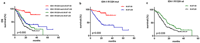
Figure 1: Kaplan–Meier survival curves and Breslow tests for IDH-1R132H mut and Ki-67≥30 in diffuse gliomas. Patient were separated into groups based on IDH-1R132H mut and Ki-67 index; clinical outcomes differed among the groups a. Ki-67<30 was associated with longer overall survival in both IDH-1R132H mut b. and IDH-1R132H wt c. patients.
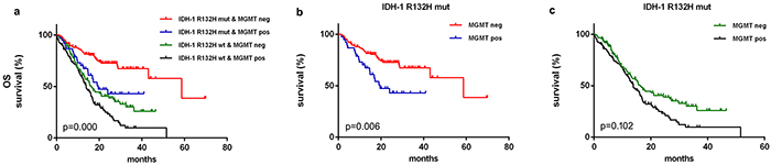
Figure 2: Kaplan–Meier survival curves and Breslow tests for IDH-1R132H mut and MGMT proteins in diffuse gliomas. Overall survival differed depending on IDH-1R132H mut status and MGMT protein levels a. Lower MGMT levels were associated with better clinical outcomes in IDH-1 R132H mut b. but not in IDH-1R132H wt c. patients.
IDH-1R132H mutation and MGMT
The mOS of 15.670 months (95% CI 13.273–18.067) in MGMT-positive patients was shorter than the mOS of 32.500 months (95% CI 22.439–42.561) observed in MGMT-negative patients (p<0.001, Breslow test). Furthermore, mOS differed among IDH-1R132H-mut-MGMTneg (58.700 months, 95% CI 31.437–85.963), IDH-1R132H mut-MGMTpos (19.570 months, 95% CI 11.018-28.122), IDH-1R132H wt-MGMTneg (16.300 months, 95% CI 13.188–19.412) and IDH-1R132H-WT-MGMTpos (13.830 months, 95% CI 11.019-16.641) patients (p<0.001, Breslow test, Figure 2a). MGMTneg patients in the IDH-1R132H mut group (p=0.006, Breslow test, Figure 2b), but not in the IDH-1R132H wt group (p=0.102, Breslow test, Figure 2c), had a much better prognosis than MGMTpos patients. No difference was observed in OS between IDH-1R132H-mut-MGMTpos and IDH-1R132H-WT-MGMTneg patients (p=0.471, Breslow test).
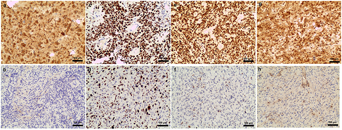
Figure 3: Examples of IDH-1R132H mut, Ki-67 index, and mutant p53 and MGMT protein levels in glioblastomas. IDH-1R132H mut positive a. and negative b. Ki-67 50%-60% c. and 20% d. mutant p53 positive e. and negative f. MGMT positive g. and negative f.
DISCUSSION
The most frequent IDH-1 mutation type in glioma is R132H, which accounts for 88.2%-92.7% of mutations in this gene [16–18]. In an IHC study specifically detecting IDH-1R132H mut using the H09 antibody, the rates were 83.0% in A II, 90.0% in O II, 100% OA II, 81.0% in A III, 88.0% in O III, 87.5% in OA III, 4% in glioblastoma, and 71.4% in sGBM [14]. The frequency of IDH-1 mutations ranged from 4-7.6% in primary glioblastoma and 73-88% in secondary glioblastoma [19, 20]. The IDH-1 R132H mutation rates detected here are similar to the ranges published previously [14, 17, 19, 21].
Also in agreement with previous findings, patients harboring IDH-1R132H mut in all grades of glioma in our study were younger than those without the mutation [6, 17, 22, 23]. Moreover, IDH-1R132H mut was present much more frequently in younger A, AA, AOA, pGBM, and sGBM patients. Other studies suggest a strong association between age and the prevalence of all WHO2007 glioma subtypes [6, 17]. In this regard, the differences between our findings and previous studies were likely due, at least in part, to small numbers of patients with particular gliomas subtypes examined here. A II IDHmut and A III IDHmut patients did not differ in age in a previous study [9]. While patients with A III were older than those with A II in our study, both groups were significantly younger than those with pGBM. However, there were no differences in patient age after they were separated based on IDH-1R132H mut. Thus, the differences in age associated with different WHO2007 grades may be strongly influenced by IDH-1R132H mut status.
Previous studies suggested a strong correlation between IDH mutations and lower Ki-67 index, mutant p53 levels, and MGMT promoter methylation in gliomas [10, 16, 19, 24–26]. Here, we found that lower Ki-67 was associated with IDHR132H mut in all WHO2007 glioma subtypes. However, mutant p53 expression in astrocytic tumors only differed after patients were grouped based on IDH-1R132H mutation status. It is well-established that IDH mutations are associated with MGMT promoter methylation [10, 26–28]. In the present study, an association between IDH-1R132H mut and MGMT protein levels was observed in glioma overall, but not within the glioma subtypes. These differences may be due to differences in the sensitivity of the detection methods [26, 29]. However, our results indicate that combining IDH-1R132H status with Ki-67 index and mutant p53 and MGMT protein levels could improve prognosis predictions in glioma patients.
Recent data suggests that IDH-1 mutations, Ki-67 index, and MGMT protein levels are prognostic factors for diffuse gliomas [15, 20, 22, 24, 30-32]. Cai et al. found that IDH-wt plus Ki-67-low and IDH-wt plus Ki-67-high astrocytic tumor patients had different clinical outcomes. The cutoff for Ki-67 in their study was 10%, and median survival was about 2 years in Ki-67-low and 1 year Ki-67-high patients [32]. Zeng et al. observed that Ki-67≥30 was associated with worse prognosis in both IDH mut (median OS=566 days) and IDH wt (median OS=355 days) groups. The median OS in our study was closer to that found by Zeng et al. These results indicate that Ki-67 index is a reliable candidate for determining prognosis in glioma patients in addition to IDH-1 status. Although the prognostic value of MGMT protein levels is controversial [29], we found here that they were predictive of prognosis. Different IHC detection thresholds may help explain this discrepancy.
In summary, we characterized the expression of IDH-1R132H mut in a large cohort of glioma patients. IDH-1R132H mut was associated with specific WHO2007 histological grades and younger age. Age differences between different WHO2007 grades of astrocytoma were strongly influenced by IDH-1R132H mutation status. Low Ki-67 index values occurred much more often in patients with lower WHO2007 grades and IDH-1R132H mutation. Finally, our study indicated that Ki-67 index and MGMT protein levels, together with IDH mutation status, were predictive of prognosis in different glioma subtypes.
materials and METHODS
Patients and tumor samples
Tumor samples were obtained from Sanbo Brain Hospital. Informed consent was obtained from all patients prior to the study. All experiments using human tissues were approved by the Institutional Review Board of Sanbo Brain Hospital. 670 adult patients with diffuse supratentorial gliomas were involved in the study. WHO classification of all specimens was performed by two independent neuropathologists [2]. In the case of a discrepancy, the two observers simultaneously reviewed the slides until a consensus was achieved. Clinical data, including patients’ age at diagnosis, sex, and molecular pathology, were collected. The diagnosis of GBMO (glioblastoma with an oligodendroglioma component) was made as previously described [33]. OS (overall survival) was measured from the date of operation to the death or the last known follow-up.
Evaluation of IDH-1R132H mut, MGMT, mutant P53, and Ki-67 levels by immunohistochemistry
Experimental procedures were performed as described previously [34–36]. Primary antibodies against IDH1R132H (Dianova 1:100), p53 (1:100 Invitrogen), MGMT (1:150 Invitrogen), and Ki-67 (1:200 Invitrogen) were used. The cutoff values were 10% for IDH-1R132H mut, 10% for mutant p53, 10% for MGMT, and 30% for Ki-67. Representative images of high and low IDH-1R132H mut (Figure 3a, 3b), Ki-67 (Figure 3c, 3d), mutant p53 (Figure 3e, 3f), and MGMT protein (Figure 3g, 3h) levels in glioblastoma patients are shown.
Statistics
SPSS 22.0 was used for all statistical analyses. The χ2 test was applied to assess the co-occurrence of IDH-1 mutation patient characteristics or the presence of other disease biomarkers. Survival curves were analyzed by the Kaplan-Meier method and the Breslow test. A p<0.05 (two-sided) was considered statistically significant.
FUNDING
This work was supported by grants from the National Youth Science Fund from China (No.81302200) and Key Projects in the National Science & Technology Pillar Program during the Twelfth Five-year Plan Period (No.2014BAI04B01) from China.
CONFLICTS OF INTEREST
The authors declare that they have no conflict of interest.
REFERENCES
1. Dolecek TA, Propp JM, Stroup NE and Kruchko C. CBTRUS statistical report: primary brain and central nervous system tumors diagnosed in the United States in 2005-2009. Neuro-oncology. 2012; 14:v1-49.
2. Louis DN, Ohgaki H, Wiestler OD, Cavenee WK, Burger PC, Jouvet A, Scheithauer BW and Kleihues P. The 2007 WHO classification of tumours of the central nervous system. Acta neuropathologica. 2007; 114:97-109.
3. Van Meir EG, Hadjipanayis CG, Norden AD, Shu HK, Wen PY and Olson JJ. Exciting new advances in neuro-oncology: the avenue to a cure for malignant glioma. CA Cancer J Clin. 2010; 60:166-193.
4. Megova M, Drabek J, Koudelakova V, Trojanec R, Kalita O and Hajduch M. Isocitrate dehydrogenase 1 and 2 mutations in gliomas. Journal of neuroscience research. 2014; 92:1611-1620.
5. Verhaak RG, Hoadley KA, Purdom E, Wang V, Qi Y, Wilkerson MD, Miller CR, Ding L, Golub T, Mesirov JP, Alexe G, Lawrence M, O’Kelly M, Tamayo P, Weir BA, Gabriel S, et al. Integrated genomic analysis identifies clinically relevant subtypes of glioblastoma characterized by abnormalities in PDGFRA, IDH1, EGFR, and NF1. Cancer cell. 2010; 17:98-110.
6. Balss J, Meyer J, Mueller W, Korshunov A, Hartmann C and von Deimling A. Analysis of the IDH1 codon 132 mutation in brain tumors. Acta neuropathologica. 2008; 116:597-602.
7. Cohen AL, Holmen SL and Colman H. IDH1 and IDH2 mutations in gliomas. Current neurology and neuroscience reports. 2013; 13:345.
8. Sanson M, Marie Y, Paris S, Idbaih A, Laffaire J, Ducray F, El Hallani S, Boisselier B, Mokhtari K, Hoang-Xuan K and Delattre JY. Isocitrate dehydrogenase 1 codon 132 mutation is an important prognostic biomarker in gliomas. Journal of clinical oncology. 2009; 27:4150-4154.
9. Reuss DE, Mamatjan Y, Schrimpf D, Capper D, Hovestadt V, Kratz A, Sahm F, Koelsche C, Korshunov A, Olar A, Hartmann C, Reijneveld JC, Wesseling P, Unterberg A, Platten M, Wick W, et al. IDH mutant diffuse and anaplastic astrocytomas have similar age at presentation and little difference in survival: a grading problem for WHO. Acta neuropathologica. 2015; 129:867-873.
10. Shibahara I, Sonoda Y, Shoji T, Kanamori M, Saito R, Inoue T, Kawaguchi T, Yamashita Y, Watanabe T, Kumabe T, Watanabe M, Suzuki H and Tominaga T. Malignant clinical features of anaplastic gliomas without IDH mutation. Neuro-oncology. 2015; 17:136-144.
11. van den Bent MJ, Hartmann C, Preusser M, Strobel T, Dubbink HJ, Kros JM, von Deimling A, Boisselier B, Sanson M, Halling KC, Diefes KL, Aldape K and Giannini C. Interlaboratory comparison of IDH mutation detection. Journal of neuro-oncology. 2013; 112:173-178.
12. Preusser M, Capper D, Hartmann C and Euro CNSRC. IDH testing in diagnostic neuropathology: review and practical guideline article invited by the Euro-CNS research committee. Clinical neuropathology. 2011; 30:217-230.
13. Reuss DE, Sahm F, Schrimpf D, Wiestler B, Capper D, Koelsche C, Schweizer L, Korshunov A, Jones DT, Hovestadt V, Mittelbronn M, Schittenhelm J, Herold-Mende C, Unterberg A, Platten M, Weller M, et al. ATRX and IDH1-R132H immunohistochemistry with subsequent copy number analysis and IDH sequencing as a basis for an “integrated” diagnostic approach for adult astrocytoma, oligodendroglioma and glioblastoma. Acta neuropathologica. 2015; 129:133-146.
14. Capper D, Reuss D, Schittenhelm J, Hartmann C, Bremer J, Sahm F, Harter PN, Jeibmann A and von Deimling A. Mutation-specific IDH1 antibody differentiates oligodendrogliomas and oligoastrocytomas from other brain tumors with oligodendroglioma-like morphology. Acta neuropathologica. 2011; 121:241-252.
15. Ogura R, Tsukamoto Y, Natsumeda M, Isogawa M, Aoki H, Kobayashi T, Yoshida S, Okamoto K, Takahashi H, Fujii Y and Kakita A. Immunohistochemical profiles of IDH1, MGMT and P53: Practical significance for prognostication of patients with diffuse gliomas. Neuropathology. 2015; 35:324-335.
16. Yan H, Parsons DW, Jin G, McLendon R, Rasheed BA, Yuan W, Kos I, Batinic-Haberle I, Jones S, Riggins GJ, Friedman H, Friedman A, Reardon D, Herndon J, Kinzler KW, Velculescu VE, et al. IDH1 and IDH2 mutations in gliomas. The New England journal of medicine. 2009; 360:765-773.
17. Hartmann C, Meyer J, Balss J, Capper D, Mueller W, Christians A, Felsberg J, Wolter M, Mawrin C, Wick W, Weller M, Herold-Mende C, Unterberg A, Jeuken JW, Wesseling P, Reifenberger G, et al. Type and frequency of IDH1 and IDH2 mutations are related to astrocytic and oligodendroglial differentiation and age: a study of 1,010 diffuse gliomas. Acta neuropathologica. 2009; 118:469-474.
18. Leeper HE, Caron AA, Decker PA, Jenkins RB, Lachance DH and Giannini C. IDH mutation, 1p19q codeletion and ATRX loss in WHO grade II gliomas. Oncotarget. 2015; 6:30295-30305. doi: 10.18632/oncotarget.4497.
19. Ohgaki H and Kleihues P. The definition of primary and secondary glioblastoma. Clinical cancer research. 2013; 19:764-772.
20. Wang XW, Ciccarino P, Rossetto M, Boisselier B, Marie Y, Desestret V, Gleize V, Mokhtari K, Sanson M and Labussiere M. IDH mutations: genotype-phenotype correlation and prognostic impact. BioMed research international. 2014; 2014:540236.
21. Capper D, Weissert S, Balss J, Habel A, Meyer J, Jager D, Ackermann U, Tessmer C, Korshunov A, Zentgraf H, Hartmann C and von Deimling A. Characterization of R132H mutation-specific IDH1 antibody binding in brain tumors. Brain pathology. 2010; 20:245-254.
22. Parsons DW, Jones S, Zhang X, Lin JC, Leary RJ, Angenendt P, Mankoo P, Carter H, Siu IM, Gallia GL, Olivi A, McLendon R, Rasheed BA, Keir S, Nikolskaya T, Nikolsky Y, et al. An integrated genomic analysis of human glioblastoma multiforme. Science. 2008; 321:1807-1812.
23. Yan W, Zhang W, You G, Bao Z, Wang Y, Liu Y, Kang C, You Y, Wang L and Jiang T. Correlation of IDH1 mutation with clinicopathologic factors and prognosis in primary glioblastoma: a report of 118 patients from China. PloS one. 2012; 7:e30339.
24. Zeng A, Hu Q, Liu Y, Wang Z, Cui X, Li R, Yan W and You Y. IDH1/2 mutation status combined with Ki-67 labeling index defines distinct prognostic groups in glioma. Oncotarget. 2015; 6: 30232-30238. doi: 10.18632/oncotarget.4920.
25. Mulholland S, Pearson DM, Hamoudi RA, Malley DS, Smith CM, Weaver JM, Jones DT, Kocialkowski S, Backlund LM, Collins VP and Ichimura K. MGMT CpG island is invariably methylated in adult astrocytic and oligodendroglial tumors with IDH1 or IDH2 mutations. International journal of cancer. 2012; 131:1104-1113.
26. Mellai M, Monzeglio O, Piazzi A, Caldera V, Annovazzi L, Cassoni P, Valente G, Cordera S, Mocellini C and Schiffer D. MGMT promoter hypermethylation and its associations with genetic alterations in a series of 350 brain tumors. Journal of neuro-oncology. 2012; 107:617-631.
27. Le Rhun E, Taillibert S and Chamberlain MC. Anaplastic glioma: current treatment and management. Expert review of neurotherapeutics. 2015:1-20.
28. Wiestler B, Capper D, Sill M, Jones DT, Hovestadt V, Sturm D, Koelsche C, Bertoni A, Schweizer L, Korshunov A, Weiss EK, Schliesser MG, Radbruch A, Herold-Mende C, Roth P, Unterberg A, et al. Integrated DNA methylation and copy-number profiling identify three clinically and biologically relevant groups of anaplastic glioma. Acta neuropathologica. 2014; 128:561-571.
29. Cankovic M, Nikiforova MN, Snuderl M, Adesina AM, Lindeman N, Wen PY and Lee EQ. The role of MGMT testing in clinical practice: a report of the association for molecular pathology. J Mol Diagn. 2013; 15:539-555.
30. Xia L, Wu B, Fu Z, Feng F, Qiao E, Li Q, Sun C and Ge M. Prognostic role of IDH mutations in gliomas: a meta-analysis of 55 observational studies. Oncotarget. 2015; 6:17354-17365. doi: 10.18632/oncotarget.4008.
31. Zhang CB, Bao ZS, Wang HJ, Yan W, Liu YW, Li MY, Zhang W, Chen L and Jiang T. Correlation of IDH1/2 mutation with clinicopathologic factors and prognosis in anaplastic gliomas: a report of 203 patients from China. J Cancer Res Clin Oncol. 2014; 140:45-51.
32. Cai J, Yang P, Zhang C, Zhang W, Liu Y, Bao Z, Liu X, Du W, Wang H, Jiang T and Jiang C. ATRX mRNA expression combined with IDH1/2 mutational status and Ki-67 expression refines the molecular classification of astrocytic tumors: evidence from the whole transcriptome sequencing of 169 samples samples. Oncotarget. 2014; 5:2551-2561. doi: 10.18632/oncotarget.1838.
33. Wang Y, Li S, Chen L, You G, Bao Z, Yan W, Shi Z, Chen Y, Yao K, Zhang W, Kang C and Jiang T. Glioblastoma with an oligodendroglioma component: distinct clinical behavior, genetic alterations, and outcome. Neuro-oncology. 2012; 14:518-525.
34. Yao K, Wang H, Duan Z, Bian Y, Xia L, Ma Z and Qi X. Mixed granular cell astrocytoma and fibrosarcoma of the brain: a case report. International journal of clinical and experimental pathology. 2014; 7:4473-4478.
35. Yao K, Wu B, Xi M, Duan Z, Wang J and Qi X. Distant dissemination of mixed low-grade astroblastoma-arteriovenous malformation after initial operation: a case report. International journal of clinical and experimental pathology. 2015; 8:7450-7456.
36. Yao K, Duan Z, Mei X, Xu Y, Li J, Li S and Qi X. A rare case of malignant pediatric ectomesenchymoma arising from the cerebrum. International journal of clinical and experimental pathology. 2015; 8:8545-8550.