
Introduction
Colorectal cancer (CRC) is a common disease. In 2021, an estimated 149,500 new cases of CRC and 52,980 of CRC-related deaths are expected [1]. The treatment of locally advanced rectal cancer is different from colon cancer. Surgical resection is the cornerstone treatment modality following a course of neoadjuvant therapy [2, 3]. Tumor response to neoadjuvant therapy may predict long term outcomes such as disease-free survival (DFS) and overall survival (OS) [4].
Exosomes are a subset of extracellular vesicles that are 40–100 nm in diameter [5]. Exosomal membranes are enriched in endosome-specific tetraspanins (CD9, CD63 and CD81) [6]. Exosomes are functional nanocarriers of a complex cargo of proteins, lipids, and nucleic acids and transfer it between the donor and recipient cells. Thus, they represent a novel mode of intercellular communication playing important roles in tumor progression, metastasis, chemotherapy resistance and immune response [5, 7–14]. Exosomes and their biologically active cargo differ between different cells and may offer prognostic information [14–16].
During treatment with chemotherapy and/or radiotherapy, exosomes and their content undergo different changes that play a role in tumor characteristics. Radiation-induced changes in exosomes promote migration of recipient cells and may facilitate progression during radiotherapy [17]. Radiation-derived exosomes promoted proliferation and helped recipient cancer cells to survive radiation in vitro and decreased the survival of tumor-bearing mice in vivo [18]. Chemotherapy-induced exosomes have been shown to carry different cargo loads compared to no-chemotherapy-induced exosomes. Apart from chemoresistance, there is growing evidence to show that chemotherapy-induced exosomes influence tumor behavior, metastasis and immune response [10, 19].
Neoadjuvant rectal (NAR) score is a short-term surrogate endpoint for predicting long-term endpoints such as OS. Calculating the NAR score is performed using tumor variables before and after treatment with neoadjuvant therapy including the clinical T (cT), the pathological T (pT) and pathological N (pN) stages using the formula ([5 ypN − 3 (cT − ypT) + 12]2/9.61) [20, 21]. The NAR score was validated using the patient dataset from NSABP R-04 clinical trial where NAR scores were categorized as low, intermediate, and high. Low (NAR < 8), intermediate (NAR = 8–16) and high (NAR > 16) scores were associated with OS (p = 0.0001) with 5-year OS of 92, 89 and 68% respectively [22].
The impact of NCCR on CD63 and CD9 expression and their prognostic significance in patients with rectal adenocarcinoma is yet to be explored. In this study, we explored the impact of NCCR on CD63 and CD9 expression pattern and their prognostic significance using the short-term surrogate endpoint NAR score.
Results
Patients baseline characteristics
Our cohort (N = 33) included patients identified between 2015 and 2018, with rectal adenocarcinoma treated with NCCR and had pre-NCCR biopsy and post-NCCR resected rectum examined for exosomal markers expression using IHC. Patients’ baseline characteristics are summarized in Table 1. The Median age was 59 years (range 34–71). Caucasians, African Americans and other ethnicities represented 23 (70%), 9 (27%) and 1 (3%) patients respectively. Males and Females represented 26 (79%) and 7 (21%) patients respectively. The primary tumor stage as assessed by pelvic magnetic resonance imaging (MRI) was cT1, cT2, cT3 and cT4 in 0 (0%), 2 (6%), 29 (88%) and 2 (6%) patients respectively. The lymph node stage as assessed by pelvic MRI was cN0, cN1 and cN2 in 10 (30%), 12 (36%) and 11 (33%) patients respectively. All but 3 (9%) patients had no evidence of distant metastasis. In the patients with distant metastasis, the site of metastasis was liver in 2 patients and peritoneum in 1 patient. Patients with low, intermediate and high NAR scores were 2 (6%), 17 (52%) and 14 (42%) patients respectively.
Table 1: Patients baseline characteristics (N = 33)
| Age (Years) | |
| Median (Range) | 59 (34–71) |
| Sex | N (%) |
| Male | 26 (79%) |
| Female | 7 (21%) |
| Ethnicity | N (%) |
| White | 23 (70%) |
| African American | 9 (27%) |
| Other | 1 (3%) |
| Stage | N (%) |
| cT stage | |
| cT1 | 0 (0%) |
| cT2 | 2 (6%) |
| cT3 | 29 (88%) |
| cT4 | 2 (6%) |
| cN stage | |
| cN0 | 10 (30%) |
| cN1 | 12 (36%) |
| cN2 | 11 (33%) |
| cM stage | |
| cM0 | 30 (91%) |
| cM1 | 3 (9%) |
| NAR score | N (%) |
| Low (<8) | 2 (6%) |
| Intermediate (= 8–16) | 17 (52%) |
| High (>16) | 14 (42%) |
Expression of CD63 and CD9 using immunohistochemistry
The expression of CD63 and CD9 using IHC in the pre-NCCR biopsy is shown in Figure 1A and 1B respectively. The expression of CD63 and CD9 using IHC in the post-NCCR resected rectum is shown in Figure 2A and 2B respectively.
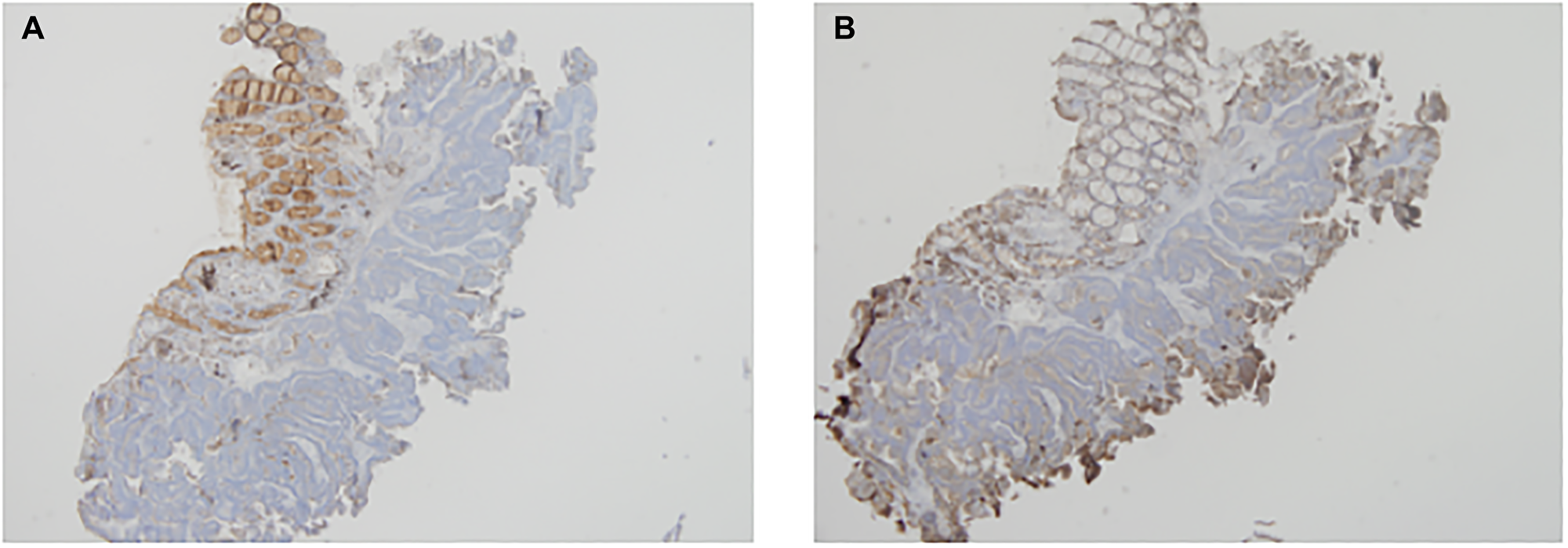
Figure 1: The expression of CD63 and CD9 using IHC in the pre-NCCR biopsy is shown in (A) and (B) respectively.
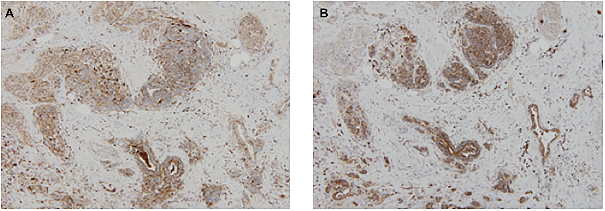
Figure 2: The expression of CD63 and CD9 using IHC in the post-NCCR resected rectum is shown in (A) and (B) respectively.
Tumor exosomal markers (CD63 and CD9) expression scores
The mean tumor CD63 score in pre-NCCR rectal biopsy vs. post-NCCR resected rectum was 106 vs. 165 (P = 0.0022). The mean tumor CD9 score in pre-NCCR rectal biopsy vs. post-NCCR resected rectum was 136 vs. 215 (p < 0.0001). The mean tumor scores for CD63 and CD9 in pre-NCCR rectal biopsy vs. post-NCCR resected rectum are summarized in Table 2. The differences in the median tumor CD63 and CD9 scores between pre-NCCR rectal biopsy and post-NCCR resected rectum is visualized in Figure 3A.
Table 2: The mean tumor and adjacent normal mucosa scores for CD63 and CD9 in pre-NCCR rectal biopsy vs. post-NCCR resected rectum
| Tissue | Number | CD63 Mean score Median (SD) | CD9 Mean score Median (SD) |
|---|---|---|---|
| Tumor | |||
| Pre-NCCR Rectal Biopsy | 33 | 106 (73.9) | 136 (66.1) |
| Post-NCCR Resected Rectum | 33 | 165 (79.6) | 215 (65.5) |
| (P = 0.0022) | (P < 0.0001) | ||
| Adjacent Normal Mucosa | |||
| Pre-NCCR Rectal Biopsy | 16 | 166 (92.5) | 104 (76.2) |
| Post-NCCR Resected Rectum | 16 | 183 (79.4) | 145 (76.1) |
| (P = 0.37) | (P = 0.0897) | ||
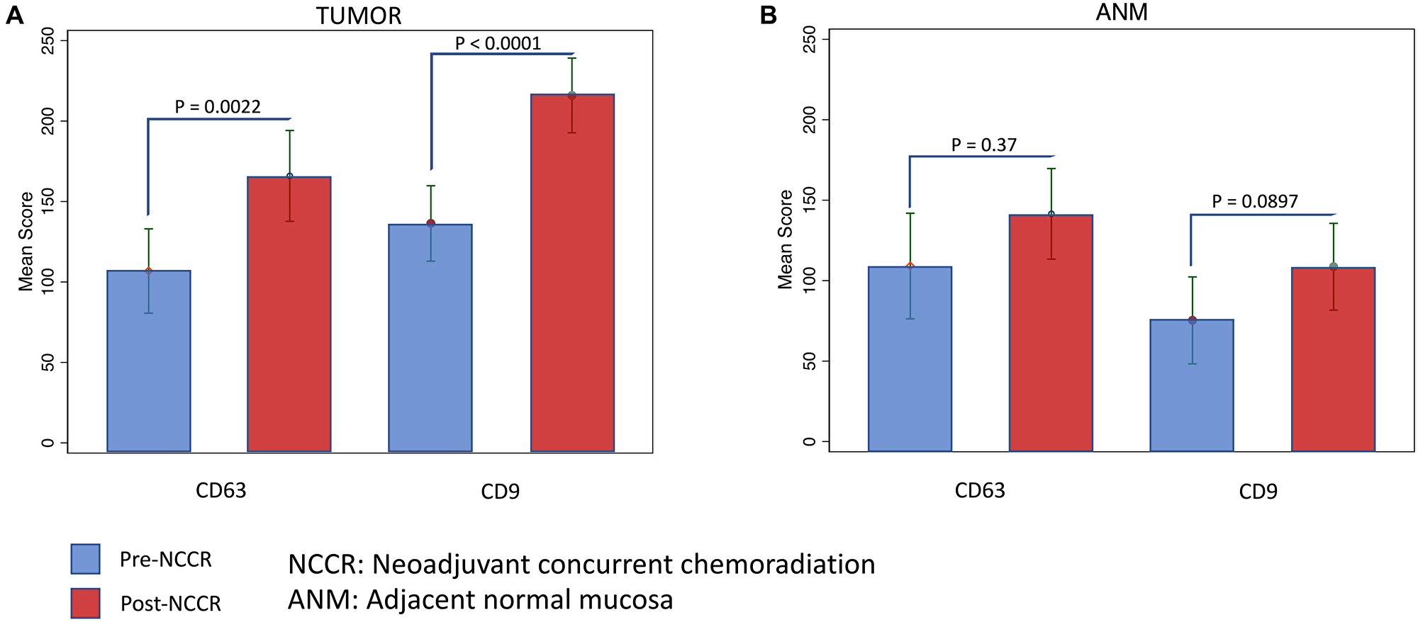
Figure 3: The differences in the median tumor and ANM CD63 and CD9 scores between pre-NCCR rectal biopsy and post-NCCR resected rectum is visualized in (A) and (B) respectively.
Adjacent normal mucosa exosomal markers (CD63 and CD9) expression scores
The exosomal markers expression scoring in the ANM from pre-NCCR rectal biopsy and post-NCCR resected rectum was only performed in 16 out of 33 patients (due to ANM tissue availability). The mean ANM CD63 score in pre-NCCR rectal biopsy vs. post-NCCR resected rectum was 166 vs. 183 (p = 0.37). The mean tumor CD9 score in pre-NCCR rectal biopsy vs. post-NCCR resected rectum was 104 vs. 145 (p = 0.0897). The mean ANM scores for CD63 and CD9 in pre-NCCR rectal biopsy vs. post-NCCR resected rectum are summarized in Table 2. The differences in the median ANM CD63 and CD9 scores between pre-NCCR rectal biopsy and post-NCCR resected rectum is visualized in Figure 3B.
The prognostic value of tumor exosomal markers (CD63 and CD9)
Only 2 patients had low NAR score, and thus low and intermediate NAR scores were grouped together. The tumor pre-NCCR rectal biopsy mean CD63 score was 99 in patients with low-intermediate NAR scores compared to 117 in patients with high NAR scores (P = 0.4934). The tumor post-NCCR resected rectum mean CD63 score was 155 in patients with low-intermediate NAR scores compared to 180 in patients with high NAR scores (P = 0.3793). The tumor pre-NCCR rectal biopsy mean CD9 score was 130 in patients with low-intermediate NAR scores compared to 144 in patients with high NAR scores (P = 0.5519). The tumor post-NCCR resected rectum mean CD9 score was 205 in patients with low-intermediate NAR scores compared to 230 in patients with high NAR scores (P = 0.2837). The mean tumor scores for CD63 and CD9 in pre-NCCR rectal biopsy and post-NCCR resected rectum according to NAR score are summarized in Table 3.
Table 3: The mean tumor scores for CD63 and CD9 in pre NCCR rectal biopsy and post NCCR resected rectum according to NAR score
| Neoadjuvant Rectal Score | Number | Tumor Pre-NCCR Mean score | Tumor Post-NCCR Mean score |
|---|---|---|---|
| Exosomal Marker CD63 | |||
| Low-Intermediate (NAR ≤ 16) | 19 | 99 | 155 |
| High (NAR > 16) | 14 | 117 | 180 |
| (P = 0.4934) | (P < 0.3793) | ||
| Exosomal Marker CD9 | |||
| Low-Intermediate (NAR ≤ 16) | 19 | 130 | 205 |
| High (NAR > 16) | 14 | 144 | 230 |
| (P = 0.5519) | (P = 0.2837) | ||
DISCUSSION
Exploring exosomes and the mechanisms that govern their generation and functions is motivated by their potential as diagnostic, therapeutic and prognostic tools in many diseases especially cancer [16, 23]. Increasingly, exosomes research is aimed at isolation methods, understanding their diverse range of biological functions and exploring their local and distant interactions [24–26]. Current characterization of biological activities of exosomes in cancer has largely been carried out on cell lines and animal models and therefore it remains unclear if some of the reported properties and functions of exosomes will be reproduced in blood or tumor specimens [27, 28].
During treatment with radiotherapy, exosomes and their content undergo different changes that play a role in tumor characteristics. Exosomes isolated from irradiated donor cells boost the motility of the head and neck squamous cell carcinoma (HNSCC) cells BHY and FaDu through enhanced AKT-signaling. That promoted migration of HNSCC cells is potentially driving HNSCC progression during treatment with radiotherapy [17]. In STS26T human malignant peripheral nerve sheath tumor cells, U87 Glioma cells and SH-SY5Y human neuroblastoma cells, radiation-derived exosomes increased proliferation and enabled recipient cancer cells to survive radiation in vitro. Moreover, radiation-derived exosomes increased tumor burden and decreased survival in an in vivo model [18]. In breast cancer cells (MCF-7), exosome biogenesis and secretion could be activated by X-ray in a dose-dependent fashion suggesting the therapeutic response of cells via ROS and exosome activity [29]. Chemotherapy-induced exosomes have been shown to carry different cargo loads compared to no-chemotherapy-induced exosomes [10]. Apart from mediating chemotherapy resistance, there is growing evidence to show that chemotherapy-induced exosomes influence tumor behavior, metastasis and immune response. [10, 19] Research efforts to explore the impact of chemotherapy concurrent with radiotherapy on exosomes are needed, and patients with rectal adenocarcinoma is a group of patients that will likely benefit from such efforts.
In our patients with rectal cancer, a differential expression of exosomal markers was observed between tumor tissue and ANM in both pre-NCCR rectal biopsies and post-NCCR resected rectum specimens (Table 2). Exposure to NCCR resulted in an increase in the tumor exosomal markers (CD63 and CD9) expression in the post-NCCR resected rectum compared to pre-NCCR rectal biopsies. There was a trend toward increased expression of ANM exosomal markers (CD63 and CD9) in the post-NCCR resected rectum compared to pre-NCCR rectal biopsies but it didn’t reach statistical significance (probably due to small sample size, N = 16). To our knowledge, our study is the first to show that exposure to NCCR increases the expression of CD63 and CD9 in patients with rectal cancer using IHC. This observation is suggestive of a possible role of exosomes in the adaptive response to NCCR in the tumor and may be the microenvironment.
Exosomal markers (CD63 and CD9) expression and its prognostic significance have been explored in different tumors. Decreased expression of CD63 and CD9 was associated with metastatic potential in patients with breast cancer, colon cancer, pancreatic cancer and Melanoma [16, 30–33]. Low expression of CD63 and CD9 was associated with poor prognosis in patients with breast cancer, pancreatic cancer, and lung cancer [16, 33, 34]. Increased CD63 expression was associated with poor prognosis in patients with gastrointestinal stromal tumors [35].
The design of clinical trials in patients with rectal adenocarcinoma has often used long-term endpoints such as DFS and OS. Unfortunately, this has slowed our progress due to the time needed for long-term outcomes to take place and for the clinical trials results to be reported. Incorporating short-term surrogate endpoints for DFS and OS such as NAR score in clinical trials design is expected to help with earlier assessment of the positive or negative interventions [20, 22].
Our work showed that the expression of CD63 and CD9 in pre-NCCR rectal biopsies and post-NCCR resected rectum specimens may have a prognostic significance. In both pre-NCCR rectal biopsies and post-NCCR resected rectum, patients with high NAR (>16) had higher CD9 and CD63 scores compared to patients with low (< 8) and intermediate NAR (8–16) scores. The lack of statistical significance is likely due to the small sample size. Overall, patients with higher CD63 and CD9 scores may have high NAR scores and worse prognoses. However, patients with lower CD63 and CD9 scores may have low-intermediate NAR scores and a better prognosis. To our knowledge, this is the first study to explore the expression pattern and prognostic significance of CD63 and CD9 in patients with rectal cancer treated with NCCR using IHC.
This study has several limitations. It included small cohort (N = 33) from a single academic cancer center. This is also a retrospective study that is prone to selection bias and provides inferior level of evidence compared to prospective studies. Moreover, the method of exosomal markers (CD63 and CD9) detection used in this study is IHC. Apart from providing data about exosomal markers expression, IHC staining doesn’t provide data about the origin of exosomes and their function and content. Despite the demonstrated impact of concurrent chemotherapy and radiation on the expression of CD63 and CD9 and their possible prognostic significance, our results and conclusion should be interpreted with caution and rather considered hypothesis-generating. Studies that can address our limitations and of larger cohorts need to be conducted to confirm the results and explore the underlying mechanisms.
Materials and Methods
Rectal cancer tissue collection
This is a retrospective study. Our cohort included patients with rectal adenocarcinoma treated with NCCR and had pre-NCCR biopsy and post-NCCR resected rectum examined for exosomal markers expression using immunohistochemistry (IHC). Patients were identified from the colorectal cancer database. This study was approved by the Institutional Review Board (IRB).
Immunohistochemical staining
Five μm sections were obtained from every rectal cancer biopsy and resected rectal cancer specimen. Two were obtained from the tumor and two were obtained from the adjacent normal mucosa (ANM). To study the expression pattern of the exosomal markers (CD63 and CD9), immunohistochemical staining was performed.
Scoring of CD63 and CD9 expression
The scoring of CD63 and CD9 staining was carried out by two pathologists. They, independently, graded the intensity of cytoplasmic staining from 1 to 3: 1 (weak), 2 (moderate) and 3 (strong) and the percentage of stained cells on each section in 10% increments. A multiplicative score was calculated for each tissue section by multiplying the percentage of positive cells by the intensity of the staining [36]. The average score between the two pathologists was calculated for each section.
Neoadjuvant rectal score calculation
Calculating the NAR score was performed using tumor variables before and after treatment with neoadjuvant therapy including the clinical T (cT), the pathological T (pT) and pathological N (pN) stages using the formula ([5 ypN − 3 (cT − ypT) + 12]2/9.61). The NAR scores were categorized as low (NAR < 8), intermediate (NAR = 8–16), and high (NAR > 16). Because only two patients had low NAR score, patients with low and intermediate NAR scores were grouped together.
Statistical analysis
The difference in CD63 and CD9 expression between pre-NCCR biopsies of the tumor/ANM and post-NCCR resected rectal specimens/ANM was compared using unpaired t-test. The difference in tumor CD63 and CD9 expression between low-intermediate and high NAR scores was compared using an unpaired t-test. Statistical significance was defined as p < 0.05, and all tests were two-sided. Tests were performed using GraphPad Prism version 9, GraphPad Software (San Diego, CA, USA), https://www.graphpad.com.
Conclusions
Using IHC, the exosomal markers (CD63 and CD9) expression increased in patients with rectal adenocarcinoma after treatment with NCCR and thus suggest a possible role of these exosomes in adaptive response to NCCR. Further follow-up and laboratory studies are required to precisely understand the underlying mechanism(s). The exosomal markers (CD63 and CD9) may have a prognostic significance. There was a trend for higher CD63 and CD9 expression in patients with high NAR score compared with low-intermediate NAR score. The lack of statistical significance is likely due to small sample size.
Abbreviations
NCCR: Neoadjuvant Concurrent Chemoradiation; IHC: Immunohistochemistry; ANM: Adjacent Normal Mucosa; CRC: Colorectal Cancer; DFS: Disease Free Survival; OS: Overall Survival; EVs: Extracellular Vesicles; IRB: Institutional Review Board; Q-score: Quick-score; HNSCC: Head and Neck Squamous Cell Carcinoma; MRI: Magnetic Resonance Imaging; NAR Score: Neoadjuvant Rectal Score.
CONFLICTS OF INTEREST
Authors have no conflicts of interest to declare.
FUNDING
This study was supported by intramural funds. Dr. Ajay P. Singh is also funded by a grant from the National Cancer Institute (5R01CA224306).
References
1. Siegel RL, Miller KD, Fuchs HE, Jemal A. Cancer Statistics, 2021. CA Cancer J Clin. 2021; 71:7–33. https://doi.org/10.3322/caac.21654. [PubMed].
2. Sauer R, Becker H, Hohenberger W, Rödel C, Wittekind C, Fietkau R, Martus P, Tschmelitsch J, Hager E, Hess CF, Karstens JH, Liersch T, Schmidberger H, Raab R, and German Rectal Cancer Study Group. Preoperative versus postoperative chemoradiotherapy for rectal cancer. N Engl J Med. 2004; 351:1731–40. https://doi.org/10.1056/NEJMoa040694. [PubMed].
3. Roh MS, Colangelo LH, O’Connell MJ, Yothers G, Deutsch M, Allegra CJ, Kahlenberg MS, Baez-Diaz L, Ursiny CS, Petrelli NJ, Wolmark N. Preoperative multimodality therapy improves disease-free survival in patients with carcinoma of the rectum: NSABP R-03. J Clin Oncol. 2009; 27:5124–30. https://doi.org/10.1200/JCO.2009.22.0467. [PubMed].
4. Das P, Skibber JM, Rodriguez-Bigas MA, Feig BW, Chang GJ, Wolff RA, Eng C, Krishnan S, Janjan NA, Crane CH. Predictors of tumor response and downstaging in patients who receive preoperative chemoradiation for rectal cancer. Cancer. 2007; 109:1750–55. https://doi.org/10.1002/cncr.22625. [PubMed].
5. Mathivanan S, Fahner CJ, Reid GE, Simpson RJ. ExoCarta 2012: database of exosomal proteins, RNA and lipids. Nucleic Acids Res. 2012; 40:D1241–44. https://doi.org/10.1093/nar/gkr828. [PubMed].
6. Taylor DD, Gercel-Taylor C. Exosomes/microvesicles: mediators of cancer-associated immunosuppressive microenvironments. Semin Immunopathol. 2011; 33:441–54. https://doi.org/10.1007/s00281-010-0234-8. [PubMed].
7. Greening DW, Gopal SK, Xu R, Simpson RJ, Chen W. Exosomes and their roles in immune regulation and cancer. Semin Cell Dev Biol. 2015; 40:72–81. https://doi.org/10.1016/j.semcdb.2015.02.009. [PubMed].
8. Gangoda L, Boukouris S, Liem M, Kalra H, Mathivanan S. Extracellular vesicles including exosomes are mediators of signal transduction: are they protective or pathogenic? Proteomics. 2015; 15:260–71. https://doi.org/10.1002/pmic.201400234. [PubMed].
9. Mittelbrunn M, Gutiérrez-Vázquez C, Villarroya-Beltri C, González S, Sánchez-Cabo F, González MÁ, Bernad A, Sánchez-Madrid F. Unidirectional transfer of microRNA-loaded exosomes from T cells to antigen-presenting cells. Nat Commun. 2011; 2:282. https://doi.org/10.1038/ncomms1285. [PubMed].
10. Patel GK, Khan MA, Bhardwaj A, Srivastava SK, Zubair H, Patton MC, Singh S, Khushman M, Singh AP. Exosomes confer chemoresistance to pancreatic cancer cells by promoting ROS detoxification and miR-155-mediated suppression of key gemcitabine-metabolising enzyme, DCK. Br J Cancer. 2017; 116:609–19. https://doi.org/10.1038/bjc.2017.18. [PubMed].
11. Simpson RJ, Lim JW, Moritz RL, Mathivanan S. Exosomes: proteomic insights and diagnostic potential. Expert Rev Proteomics. 2009; 6:267–83. https://doi.org/10.1586/epr.09.17. [PubMed].
12. Vidal M, Sainte-Marie J, Philippot JR, Bienvenue A. Asymmetric distribution of phospholipids in the membrane of vesicles released during in vitro maturation of guinea pig reticulocytes: evidence precluding a role for “aminophospholipid translocase”. J Cell Physiol. 1989; 140:455–62. https://doi.org/10.1002/jcp.1041400308. [PubMed].
13. Valadi H, Ekström K, Bossios A, Sjöstrand M, Lee JJ, Lötvall JO. Exosome-mediated transfer of mRNAs and microRNAs is a novel mechanism of genetic exchange between cells. Nat Cell Biol. 2007; 9:654–59. https://doi.org/10.1038/ncb1596. [PubMed].
14. Patel GK, Patton MC, Singh S, Khushman M, Singh AP. Pancreatic Cancer Exosomes: Shedding Off for a Meaningful Journey. Pancreat Disord Ther. 2016; 6:e148. https://doi.org/10.4172/2165-7092.1000e148. [PubMed].
15. Khushman M, Bhardwaj A, Patel GK, Laurini JA, Roveda K, Tan MC, Patton MC, Singh S, Taylor W, Singh AP. Exosomal Markers (CD63 and CD9) Expression Pattern Using Immunohistochemistry in Resected Malignant and Nonmalignant Pancreatic Specimens. Pancreas. 2017; 46:782–88. https://doi.org/10.1097/MPA.0000000000000847. [PubMed].
16. Khushman M, Patel GK, Laurini JA, Bhardwaj A, Roveda K, Donnell R, Sherling K, Case B, Frankel AE, Pai S, Taylor W, Tan MCB, Mizrahi M, et al. Exosomal markers (CD63 and CD9) expression and their prognostic significance using immunohistochemistry in patients with pancreatic ductal adenocarcinoma. J Gastrointest Oncol. 2019; 10:695–702. https://doi.org/10.21037/jgo.2018.07.02. [PubMed].
17. Mutschelknaus L, Azimzadeh O, Heider T, Winkler K, Vetter M, Kell R, Tapio S, Merl-Pham J, Huber SM, Edalat L, Radulović V, Anastasov N, Atkinson MJ, Moertl S. Radiation alters the cargo of exosomes released from squamous head and neck cancer cells to promote migration of recipient cells. Sci Rep. 2017; 7:12423. https://doi.org/10.1038/s41598-017-12403-6. [PubMed].
18. Mrowczynski OD, Madhankumar AB, Sundstrom JM, Zhao Y, Kawasawa YI, Slagle-Webb B, Mau C, Payne RA, Rizk EB, Zacharia BE, Connor JR. Exosomes impact survival to radiation exposure in cell line models of nervous system cancer. Oncotarget. 2018; 9:36083–101. https://doi.org/10.18632/oncotarget.26300. [PubMed].
19. Ab Razak NS, Ab Mutalib NS, Mohtar MA, Abu N. Impact of Chemotherapy on Extracellular Vesicles: Understanding the Chemo-EVs. Front Oncol. 2019; 9:1113. https://doi.org/10.3389/fonc.2019.01113. [PubMed].
20. Valentini V, van Stiphout RG, Lammering G, Gambacorta MA, Barba MC, Bebenek M, Bonnetain F, Bosset JF, Bujko K, Cionini L, Gerard JP, Rödel C, Sainato A, et al. Nomograms for predicting local recurrence, distant metastases, and overall survival for patients with locally advanced rectal cancer on the basis of European randomized clinical trials. J Clin Oncol. 2011; 29:3163–72. https://doi.org/10.1200/JCO.2010.33.1595. [PubMed].
21. Fahmawi Y, Smith C, Grimm L, Khullar S, Rider P, Hunter J, Iliff G, Mneimneh W, Roveda K, Wang B, Prodduturvar P, Alkharabsheh O, McCormick B, et al. Usefulness of Restaging Pelvis Magnetic Resonance Imaging After Neoadjuvant Concurrent Chemoradiotherapy in Patients With Locally Advanced Rectal Cancer. Clin Colorectal Cancer. 2020; 19:e281–87. https://doi.org/10.1016/j.clcc.2020.06.006. [PubMed].
22. George TJ Jr, Allegra CJ, Yothers G. Neoadjuvant Rectal (NAR) Score: a New Surrogate Endpoint in Rectal Cancer Clinical Trials. Curr Colorectal Cancer Rep. 2015; 11:275–80. https://doi.org/10.1007/s11888-015-0285-2. [PubMed].
23. Kanchanapally R, Khan MA, Deshmukh SK, Srivastava SK, Khushman M, Singh S, Singh AP. Exosomal Formulation Escalates Cellular Uptake of Honokiol Leading to the Enhancement of Its Antitumor Efficacy. ACS Omega. 2020; 5:23299–307. https://doi.org/10.1021/acsomega.0c03136. [PubMed].
24. Kalluri R. The biology and function of exosomes in cancer. J Clin Invest. 2016; 126:1208–15. https://doi.org/10.1172/JCI81135. [PubMed].
25. Kahlert C, Kalluri R. Exosomes in tumor microenvironment influence cancer progression and metastasis. J Mol Med (Berl). 2013; 91:431–37. https://doi.org/10.1007/s00109-013-1020-6. [PubMed].
26. Patel GK, Khan MA, Zubair H, Srivastava SK, Khushman M, Singh S, Singh AP. Comparative analysis of exosome isolation methods using culture supernatant for optimum yield, purity and downstream applications. Sci Rep. 2019; 9:5335. https://doi.org/10.1038/s41598-019-41800-2. [PubMed].
27. Mathieu M, Martin-Jaular L, Lavieu G, Théry C. Specificities of secretion and uptake of exosomes and other extracellular vesicles for cell-to-cell communication. Nat Cell Biol. 2019; 21:9–17. https://doi.org/10.1038/s41556-018-0250-9. [PubMed].
28. Harding C, Heuser J, Stahl P. Receptor-mediated endocytosis of transferrin and recycling of the transferrin receptor in rat reticulocytes. J Cell Biol. 1983; 97:329–39. https://doi.org/10.1083/jcb.97.2.329. [PubMed].
29. Jabbari N, Nawaz M, Rezaie J. Ionizing Radiation Increases the Activity of Exosomal Secretory Pathway in MCF-7 Human Breast Cancer Cells: A Possible Way to Communicate Resistance against Radiotherapy. Int J Mol Sci. 2019; 20:3649. https://doi.org/10.3390/ijms20153649. [PubMed].
30. Miyake M, Nakano K, Ieki Y, Adachi M, Huang CL, Itoi S, Koh T, Taki T. Motility related protein 1 (MRP-1/CD9) expression: inverse correlation with metastases in breast cancer. Cancer Res. 1995; 55:4127–31. [PubMed].
31. Cajot JF, Sordat I, Silvestre T, Sordat B. Differential display cloning identifies motility-related protein (MRP1/CD9) as highly expressed in primary compared to metastatic human colon carcinoma cells. Cancer Res. 1997; 57:2593–97. [PubMed].
32. Hotta H, Ross AH, Huebner K, Isobe M, Wendeborn S, Chao MV, Ricciardi RP, Tsujimoto Y, Croce CM, Koprowski H. Molecular cloning and characterization of an antigen associated with early stages of melanoma tumor progression. Cancer Res. 1988; 48:2955–62. [PubMed].
33. Sho M, Adachi M, Taki T, Hashida H, Konishi T, Huang CL, Ikeda N, Nakajima Y, Kanehiro H, Hisanaga M, Nakano H, Miyake M. Transmembrane 4 superfamily as a prognostic factor in pancreatic cancer. Int J Cancer. 1998; 79:509–16. https://doi.org/10.1002/(sici)1097-0215(19981023)79:5<509::aid-ijc11>3.0.co;2-x. [PubMed].
34. Higashiyama M, Taki T, Ieki Y, Adachi M, Huang CL, Koh T, Kodama K, Doi O, Miyake M. Reduced motility related protein-1 (MRP-1/CD9) gene expression as a factor of poor prognosis in non-small cell lung cancer. Cancer Res. 1995; 55:6040–44. [PubMed].
35. Lewitowicz P, Matykiewicz J, Koziel D, Chrapek M, Horecka-Lewitowicz A, Gluszek S. CD63 and GLUT-1 Overexpression Could Predict a Poor Clinical Outcome in GIST: A Study of 54 Cases with Follow-Up. Gastroenterol Res Pract. 2016; 2016:6478374. https://doi.org/10.1155/2016/6478374. [PubMed].
36. Detre S, Saclani Jotti G, Dowsett M. A “quickscore” method for immunohistochemical semiquantitation: validation for oestrogen receptor in breast carcinomas. J Clin Pathol. 1995; 48:876–78. https://doi.org/10.1136/jcp.48.9.876. [PubMed].