
INTRODUCTION
Bladder cancer is the ninth most common cancer worldwide [1]. It was estimated that 76,960 new cases (4.6% of all new cancer cases) and 16,390 deaths would occur due to bladder cancer in the United States in 2016 [2]. High-grade invasive bladder cancer usually has a worse prognosis than low-grade superficial tumors. Unfortunately, approximately 30% newly diagnosed bladder cancer patients have invasive cancer [3], and 10-20% of superficial tumors will progress to invasive diseases. Thus, prediction of the progression of bladder cancer in certain patients remains a major challenge. Invasive cystoscopy is the most common method to detect suspected bladder cancer, because of the low sensitivity of other diagnostic tests, like urine cytology [3]. Bladder cancer has different biological characteristics, and patients with the same disease stage may have divergent clinical courses and different outcomes after receiving the same therapy [4]. Therefore, there is an urgent need for more sensitive and specific biomarkers that provide reliable information for tumor diagnosis, and elucidate the biological behavior of these tumors. This may allow more precise assessment of and better-targeted effective therapy for bladder cancer. A number of biomarkers such as survivin [5], fascin [6], MCT1, MCT4, and CD147 [7] were found to be involved in the development and progression of bladder cancer. Among these biomarkers, CD147, or extracellular matrix metalloproteinase inducer (EMMPRIN), is a transmembrane protein that acts as an important mediator of tumor cell invasion [8]. It is overexpressed in various tumors such as breast, lung, oral, esophageal, laryngeal, and renal cancers [9–14]. In vitro suppression of CD147 has been shown to inhibit the proliferation, migration, and invasion of T24 bladder cancer cells [15]. There are also a number of studies on CD147 expression profiles in bladder cancer patients, which indicate that CD147 may serve as a prognostic biomarker. However, some of these study results are controversial. For example, some studies showed that the positive expression of CD147 predicts poor overall survival (OS) [4, 7, 15-18], while Afonso et al. [16] and Choi et al. [7] declared no significance of positive or negative expression of CD147. Furthermore, Afonso et al. [16] and Han et al. [19] claimed that CD147 predicts poor disease specific survival (DSS), which contradicts the results of Hemdan et al. [17]. Some studies showed a significant association between CD147 over-expression and tumor stage [7, 20], while others disputed this [21]. The same conflict was also seen in the studies of lymphatic invasion status [7, 22], TNM stage [21, 23], and recurrence status [18, 24]. Therefore, we conducted this meta-analysis to quantitatively evaluate the relationship between CD147 and clinicopathological features and survival of bladder cancer patients.
RESULTS
Literature search and characteristics of included studies
A total of 147 studies were identified, of which 63 were excluded because of duplication. After reading the titles and abstracts, a further 50 studies were excluded. The remaining 34 full text studies were carefully reviewed (animal studies [n=3]; review and meta-analysis [n=5]; no control group [n=2]). Finally, 24 studies [4, 7, 15-36] were included for quantitative analysis (Figure 1).
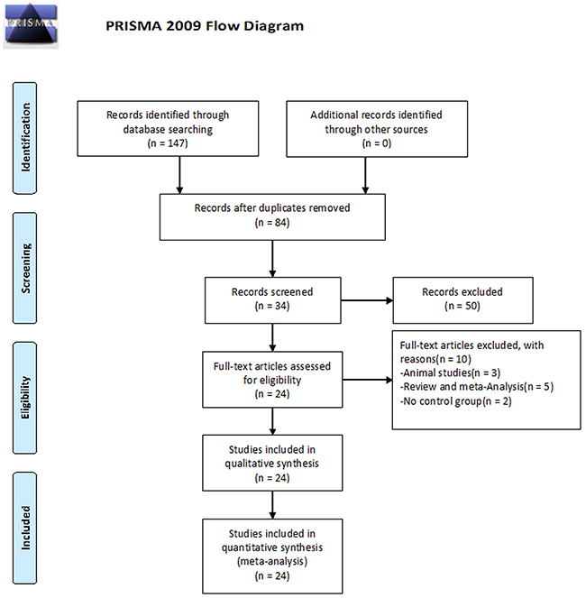
Figure 1: Flowchart of selection of studies for inclusion in meta-analysis. A total of 147 studies were identified, and 63 studies were excluded because of duplication. After reading the titles and abstracts, 50 studies were excluded. 34 possible full text studies were carefully reviewed (animal studies [n = 3]; review and meta-analysis [n = 5]; no control group [n = 2]). Finally, 24 studies were included for quantitative analysis
The characteristics of these studies are summarized in Table 1. A total of 2493 patients, with or without bladder cancer, are included in 24 studies mostly in China (sixteen studies conducted in China [15, 18, 19, 21-25, 28-35], one from Portugal [16], one from Sweden [17], one from Korea [7], one from Egypt [20], one from India [26], one from Germany [27], one from Denmark [4], and one from Japan [36]). 8 studies [4, 7, 15-19, 22] with 9 datasets (the study by Hemdan et al. [17] had 2 datasets, grouped based on differences in treatment) are included in the survival analysis (Table 2). The pathological types of bladder cancer were transitional cell carcinoma, squamous cell carcinoma, and adenocarcinoma. The studies were published between 1993 and 2015. Immunohistochemistry (IHC) was used to detect the expression of CD147 in all studies, while reverse transcriptase polymerase chain reaction, Western blot, array analysis, immunoelectron microscopy, and nucleic acid in situ hybridization were also used in six studies [4, 21, 22, 26, 27, 36].
Table 1: Characteristics of eligible studies.
First author |
Year |
Origin |
Cases |
Median age |
Counting method |
Definition of CD147 positive |
NOS score |
|---|---|---|---|---|---|---|---|
Afonso, J [16] |
2015 |
Portugal |
114 |
70 |
IHC (SABC) |
A1+B1≥4 |
8 |
Hemdan, T [17] |
2015 |
Sweden |
250 |
- |
IHC (LP) |
- |
7 |
Li, F [25] |
2015 |
China |
66 |
Case, 64.28 Control, 63.47 |
IHC (SP) |
A≥26% |
7 |
Choi, JW [7] |
2014 |
Korea |
360 |
69 |
IHC (EnVision) |
A2+B2≥16 |
7 |
Min, L [22] |
2014 |
China |
128 |
Case, 58.1 Control, 55.3 |
IHC (SP) |
A>0% or weak intensity |
7 |
El-Rehim, DM [20] |
2013 |
Egypt |
125 |
56 |
IHC (SABC) |
A3*B1>2 |
7 |
Bhagirath, D [26] |
2012 |
India |
60 |
Case, 58 Control (Benign Prostatic Hyperplasia), 50.3 Control (Healthy), 48 |
ELISA |
- |
8 |
Wittschieber, D [27] |
2011 |
Germany |
103 |
71 |
IHC (SP) |
B1≥2 |
7 |
Xue, YJ [15] |
2011 |
China |
128 |
Case, 58.3 |
IHC (SP) |
B>0 |
8 |
Gao, LJ [24] |
2011 |
China |
50 |
68 |
IHC (SABC) |
A4+B3>0 |
7 |
Li, M [21] |
2011 |
China |
79 |
60.5 |
IHC (SP) |
A≥25% |
7 |
Han, ZD [19] |
2010 |
China |
58 |
56.8 |
IHC (ABC) |
A≥6% |
8 |
Zhong, WD [18] |
2010 |
China |
131 |
Case, 68.1 |
IHC (EnVision) |
A>5% |
8 |
Chen, QB [28] |
2010 |
China |
93 |
Case, 71.5 |
IHC (SP) |
A5*B1>1 |
7 |
Cui, W [29] |
2010 |
China |
50 |
Case, 61.8 |
IHC (SP) |
A>0% |
7 |
He, HC [30] |
2009 |
China |
103 |
Case, 61.5 |
IHC (SP) |
A5*B1>1 |
7 |
Peng, XH [23] |
2009 |
China |
64 |
Case, 59.9 |
IHC (SP) |
A>0% |
7 |
Lin, JX [31] |
2008 |
China |
84 |
Case, 61.5 Control, 56.9 |
IHC (SP) |
A>0% |
7 |
Als, AB [4] |
2007 |
Denmark |
124 |
- |
IHC (SP) |
A≥10% |
8 |
Gao, L [32] |
2007 |
China |
59 |
Case, 57.8 |
IHC (SP) |
A>25% or B>0 |
7 |
Li, M [33] |
2007 |
China |
84 |
Case, 61.5 Control, 56.9 |
IHC (SP) |
A≥25% |
7 |
Li, WL [34] |
2007 |
China |
68 |
62 |
IHC (SP) |
A≥25% or B>0 |
7 |
Han, JL [35] |
2003 |
China |
54 |
61.6 |
IHC (SP) |
A≥25% or B>0 |
7 |
Muraoka, K [36] |
1993 |
Japan |
58 |
- |
IHC (ABC) |
A>30% |
7 |
Streptavidin-perosidase (SP), Labeled- perosidase (LP), Avidin-biotin complex (ABC), Streptavidin-avidin-biotin complex (SABC)
A:Positive cell percentage
A1: scored 0 (0 %), 1 (<5 %), 2 (5–50 %), 3 (>50 %).
A2: scored 0-10.
A3: scored 1 (<25 %), 2 (25–50%), 3 (51–75%), 4 (>75%).
A4: scored 0 (<10%), 1 (10-50 %), 2 (>50%).
A5: scored 0 (≤5%), 1 (6-25%), 2 (26-50%), 3 (≥50%)
B: Staining intensity
B1: scored 0 (absence of staining), 1 (weak staining), 2 (moderate staining), 3 (strong staining).
B2: scored 0-10.
B3: scored 0 (absence of staining), 1 (pale brown), 2 (dark brown)
Table 2: Characteristics of the included studies
First author |
Time |
Country |
Cases |
Tumor types |
Test method |
Cut-off value(positive) |
Follow-up time(months) |
Survival results |
HR with 95%CI |
Source of data |
|---|---|---|---|---|---|---|---|---|---|---|
Afonso, J [16] |
2015 |
Portugal |
114 |
Bladder cancer |
IHC (SABC) |
A1+B≥4 |
1-132 |
OS, DFS |
OS(U), 4.1(1.27,13.18) |
Curve + p-value |
Hemdan, T [17] |
2015 |
Sweden |
250 |
Bladder cancer |
IHC (LP) |
- |
- |
OS, DSS |
OS#(U), 3.11(1.19,8.09) |
Curve + Direct |
Choi, JW [7] |
2014 |
Korea |
360 |
Bladder cancer |
IHC (EnVision) |
A2+B2≥16 |
median, 36 |
OS |
OS(U), 2.58(0.84,7.98) |
Curve |
Min, L [22] |
2014 |
China |
128 |
Bladder cancer |
IHC (SP) |
A>0% or weak intensity |
11-86 |
DSS |
DSS(U), 3.14(1.12,8.76) |
Curve + Direct |
Xue, YJ [15] |
2011 |
China |
86 |
Bladder cancer |
IHC (SP) |
B>0 |
3-86 |
OS |
OS(U), 3.783(1.935,7.395) |
Direct |
Han, ZD [19] |
2010 |
China |
58 |
Bladder cancer |
IHC (ABC) |
A≥6% |
12-60 |
DSS |
DSS(U), 3.08(1.11,8.49) |
Curve + p-value |
Zhong, WD [18] |
2010 |
China |
131 |
Bladder cancer |
IHC (EnVision) |
A>5% |
36 |
OS, DFS |
OS(U), 2.42(1.29,4.54) |
Curve + Direct |
Als, AB [4] |
2007 |
Denmark |
124 |
Bladder cancer |
IHC (SP) |
A≥10% |
15-60 |
OS |
OS(U), 3.93(1.74,8.89) |
Indirect |
U: univariate analysis.
M: multivariate analysis.
A:Positive cell percentage
A1: scored 0 (0 %), 1 (<5 %), 2 (5–50 %), 3 (>50 %).
A2: scored 0-10.
A3: scored 1 (<25 %), 2 (25–50%), 3 (51–75%), 4 (>75%).
A4: scored 0 (<10%), 1 (10-50 %), 2 (>50%).
A5: scored 0 (≤5%), 1 (6-25%), 2 (26-50%), 3 (≥50%)
B: Staining intensity
B1: scored 0 (absence of staining), 1 (weak staining), 2 (moderate staining), 3 (strong staining).
B2: scored 0-10.
B3: scored 0 (absence of staining), 1 (pale brown), 2 (dark brown)
#: chemotherapy prior to cystectomy
*: cystectomy
Qualitative assessment
The study quality was assessed using the Newcastle-Ottawa quality assessment scale (NOS), generating scores ranging from 7 to 8 (with a mean of 7.25), where a higher value (0-9) indicates better methodology. The results of the quality assessment are shown in Table 1, with detailed information shown in Supplementary Table 1.
CD147 expression and survival analysis
CD147 expression and OS
A univariate analysis of OS was performed in 6 studies [4, 7, 15-18], including seven datasets. Without heterogeneity (P=0.349, I2=10.5%), fixed-effects model showed that the CD147-positive group had a lower OS (HR=2.63, 95% CI=[1.96, 3.53], P<0.00001). In addition, a multivariate analysis of OS was performed in 3 studies [15, 17, 18], without heterogeneity (P=0.482, I2=0%), and a fixed-effects model was used. The result was the same (HR=1.86, 95% CI=[1.32, 2.62], P=0.00036) (Figure 2A, 2B).
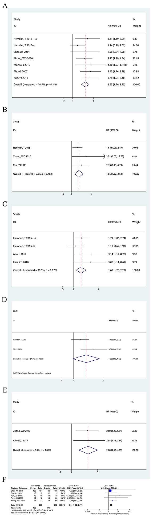
Figure 2: Forest plots of CD147 expression and survival analysis. The squares and horizontal lines correspond to the study-specific OR and 95% CI. The area of the squares reflects the study-specific weight (inverse of the variance). The diamonds represent the pooled OR/HR and 95% CI. The solid vertical line is at the null value (OR/HR=1). (A) The relationship between CD147 expression and overall survival (for univariate data; Hedman, T 2015 a: data with neoadjuvant chemotherapy and cystectomy as the therapeutic regimen, Hemdan, T 2015 b: data with only cystectomy as the therapeutic regimen). CD147 expression was associated with overall survival (HR=2.63, 95%CI= [1.96, 3.53], P<0.00001). (B) The relationship between CD147 expression and overall survival (for multivariate data). CD147 expression was associated with overall survival (HR=1.86, 95%CI= [1.32, 2.62], P=0.00036). (C) The relationship between CD147 expression and disease specific survival (for univariate data; Hedman, T 2015 a: data with neoadjuvant chemotherapy and cystectomy as the therapeutic regimen, Hemdan, T 2015 b: data with only cystectomy as the therapeutic regimen). CD147 expression was associated with disease specific survival (HR=1.65, 95%CI= [1.20, 2.27], P=0.002). (D) The relationship between CD147 expression and disease specific survival (for multivariate data). CD147 expression was associated with disease specific survival (HR=1.98, 95%CI= [0.95, 4.12], P=0.067). (E) The relationship between CD147 expression and disease recurrence-free survival (for univariate data). CD147 expression was associated with disease specific survival (HR=2.78, 95%CI= [1.56, 4.99], P=0.001). (F) The relationship between CD147 expression and recurrence. CD147 expression was associated with recurrence (OR=1.91, 95%CI= [1.32, 2.77], P=0.0006).
CD147 expression and DSS
Three studies [17, 19, 22], including four datasets, showed DSS with a univariate analysis. Without heterogeneity (P=0.175, I2=39.5%), fixed-effects model showed that the CD147-positive group had lower OS (HR=1.65, 95% CI=[1.20, 2.27], P=0.002). In addition, Hemdan et al. [17] and Min et al. [22] reported a multivariate analysis with a random-effects model, showing no difference between CD147-positive and -negative group survival (HR=1.98, 95% CI=[0.95, 4.12], P=0.067), with heterogeneity (P=0.092, I2=64.7%) (Figure 2C, 2D).
CD147 expression and DFS
Zhong et al. [18] and Afonso et al. [16] performed a univariate analysis for disease recurrence-free survival (DFS); however only Zhong et al. [18] performed a multivariate analysis, which included 101 patients, with a 3-year follow-up. Fixed-effects model showed that the CD147-positive group had poorer DFS survival (univariate analysis, P=0.864, I2=0%, HR=2.78, 95% CI=[1.56, 4.99], P=0.001; multivariate analysis, HR=5.51, 95% CI=[1.36, 22.32], P=0.017) (Figure 2E).
CD147 and recurrence
Five studies [7, 18, 23, 24, 35], utilizing 557 tissue samples, investigated the relationship of CD147 expression with the recurrence of bladder cancer. Without heterogeneity (P=0.38, I2=4%), the fixed-effects model showed a significant difference between the recurrence group and the no-recurrence group (OR=1.91, 95% CI=[1.32, 2.77], P=0.0006) (Figure 2F).
Subgroup analysis of survival data
We conducted a subgroup analysis based on IHC and cut-off values. This revealed that CD147 expression is correlated with a poor prognosis for bladder cancer in almost all subgroups (P<0.05), except multivariate analysis of OS (P=0.081) and DSS (P=0.068) in positive cell percentage only subgroup and multivariate analysis of DSS (P=0.153) in non- streptavidin-perosidase (SP) subgroup. This may attributes to the insufficiency of sample size and studies. In summary, our results are reliable (Table 3).
Table 3: Results of subgroup analysis of survival data
Based on methods of IHC |
Based on cut-off value |
|||||||
|---|---|---|---|---|---|---|---|---|
Subgroups (datasets) |
HR&OR |
95%CI |
P value |
Subgroups (datasets) |
HR&OR |
95%CI |
P value |
|
OS (U) |
SP (2) |
3.842 |
2.289-6.448 |
<0.001 |
Based on positive cell percentage only (1) |
3.93 |
1.739-8.883 |
0.01 |
Non-SP (5) |
2.203 |
1.545-3.142 |
<0.001 |
Others (6) |
2.480 |
1.812-3.394 |
<0.001 |
|
OS (M) |
SP (1) |
2.332 |
1.149-4.734 |
0.019 |
Based on positive cell percentage only (1) |
3.310 |
0.863-12.699 |
0.081 |
Non-SP (2) |
1.740 |
1.178-2.571 |
0.005 |
Others (2) |
1.790 |
1.257-2.550 |
0.001 |
|
DSS (U) |
SP (1) |
3.140 |
1.123-8.782 |
0.029 |
Based on positive cell percentage only (1) |
3.080 |
1.114-8.518 |
0.030 |
Non-SP (3) |
1.543 |
1.106-2.153 |
0.011 |
Others (3) |
1.544 |
1.106-2.155 |
0.011 |
|
DSS (M) |
SP (1) |
3.035 |
1.462-6.301 |
0.003 |
Based on positive cell percentage only (0) |
- |
- |
- |
Non-SP (1) |
1.428 |
0.876-2.328 |
0.153 |
Others (2) |
1.979 |
0.951-4.114 |
0.068 |
|
DFS (U) |
SP (0) |
- |
- |
- |
Based on positive cell percentage only (1) |
2.68 |
1.29-5.54 |
0.008 |
Non-SP (2) |
2.785 |
1.556-4.985 |
0.001 |
Others (1) |
2.98 |
1.13-7.84 |
0.027 |
|
Recurrence |
SP (2) |
11.08 |
1.35-90.77 |
0.02 |
Based on positive cell percentage only (2) |
3.14 |
1.21-8.14 |
0.02 |
Non-SP (3) |
1.73 |
1.18-2.52 |
0.005 |
Others (3) |
1.73 |
1.15-2.58 |
0.008 |
|
Overall survival (OS), Disease specific survival(DSS), Disease recurrence-free survival (DFS), univariate analysis (U), Streptavidin-perosidase (SP), multivariate analysis (M).
Non-SP includes labeled- perosidase (LP), avidin-biotin complex (ABC), streptavidin-avidin-biotin complex (SABC) and EnVision.
Others are based on staining intensity or both staining intensity and positive cell percentage.
OS (U), OS (M), DSS (U), DSS (M) and DFS (U) used HR as effect size and recurrence used OR as effect size.
CD147 expression in different bladder tissues
CD147 in bladder cancer and non-cancerous tissues
The positive expression of CD147 in bladder cancer and non-cancerous tissues was investigated in 13 studies [21-25, 28-31, 33-36] with 979 patients. Without heterogeneity (P=0.88, I2=0%), the fixed-effects model showed that CD147 expression was higher in bladder cancer tissues (OR=43.64, 95% CI=[23.26, 81.87], P<0.00001) (Figure 3A).
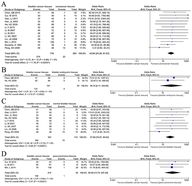
Figure 3: Forest plots of CD147 expression in different types of bladder tissues. The squares and horizontal lines correspond to the study-specific OR and 95% CI. The area of the squares reflects the study-specific weight (inverse of the variance). The diamonds represent the pooled OR and 95% CI. The solid vertical line is at the null value (OR=1). (A) CD147 positive expression between bladder cancer tissues and non-cancer bladder tissues. Significant difference was found between two groups (OR=43.64, 95%CI=[23.26, 81.87], P<0.00001). (B) CD147 positive expression between bladder cancer tissues and bladder embryonic tissues. Significant difference was found between two groups (OR=46.78, 95%CI=[6.17, 354.65], P=0.0002). (C) CD147 positive expression between bladder cancer tissues and normal bladder tissues. Significant difference was found between two groups (OR=37.99, 95%CI=[16.97, 85.03], P<0.00001). (D) CD147 positive expression between bladder cancer tissues and para-carcinoma tissues. Significant difference was found between two groups (OR=59.50, 95%CI=[13.75, 257.42], P<0.00001).
CD147 expression in bladder cancer and bladder embryonic tissues
Two studies [28, 30] reported the positive expression of CD147 in bladder cancer tissues and bladder embryonic tissues, including 147 bladder cancer tissues and 20 bladder embryonic tissues. Without heterogeneity (P=0.603, I2=0%), the fixed-effects model showed that positive expression of CD147 in bladder cancer tissues was higher than in bladder embryonic tissues (OR=46.78, 95% CI=[6.17, 354.65], P=0.0002) (Figure 3B).
CD147 expression in bladder cancer and normal bladder tissues
Nine studies [23-25, 28, 30, 31, 33-35] investigated the positive expression of CD147 in bladder cancer tissues and normal bladder tissues, including 531 bladder cancer tissues and 113 normal bladder tissues. Fixed-effects model showed that CD147 positive expression was greater in bladder cancer tissues (OR=37.99, 95% CI=[16.97, 85.03], P<0.00001), without heterogeneity (P=0.73, I2=0%) (Figure 3C).
CD147 in bladder cancer and para-carcinoma tissues
Three studies [21, 22, 29] reported the expression of CD147 in bladder cancer tissues and para-carcinoma tissues, including 215 bladder cancer tissues and 42 para-carcinoma tissues. Fixed-effects model showed a higher rate of CD147 expression in the bladder cancer group (OR=59.50, 95% CI=[13.75, 257.42], P<0.00001), without heterogeneity (P=0.94, I2 =0%) (Figure 3D).
Correlation of CD147 expression with clinicopathological parameters
CD147 and clinical stage
Clinical stage is an international standard for tumor staging. Stages are grouped into two categories, with TNM I-II having a better prognosis than TNM III-IV. The association between CD147 and clinical stage was investigated in two studies [19, 28], which showed that CD147 expression in the TNM III-IV stage group was greater than that in TNM I-II stage group, with fixed-effects model (OR=73.89, 95% CI=[9.61, 568.15], P<0.0001) and without heterogeneity (P=1.00, I2=0%) (Figure 4A).
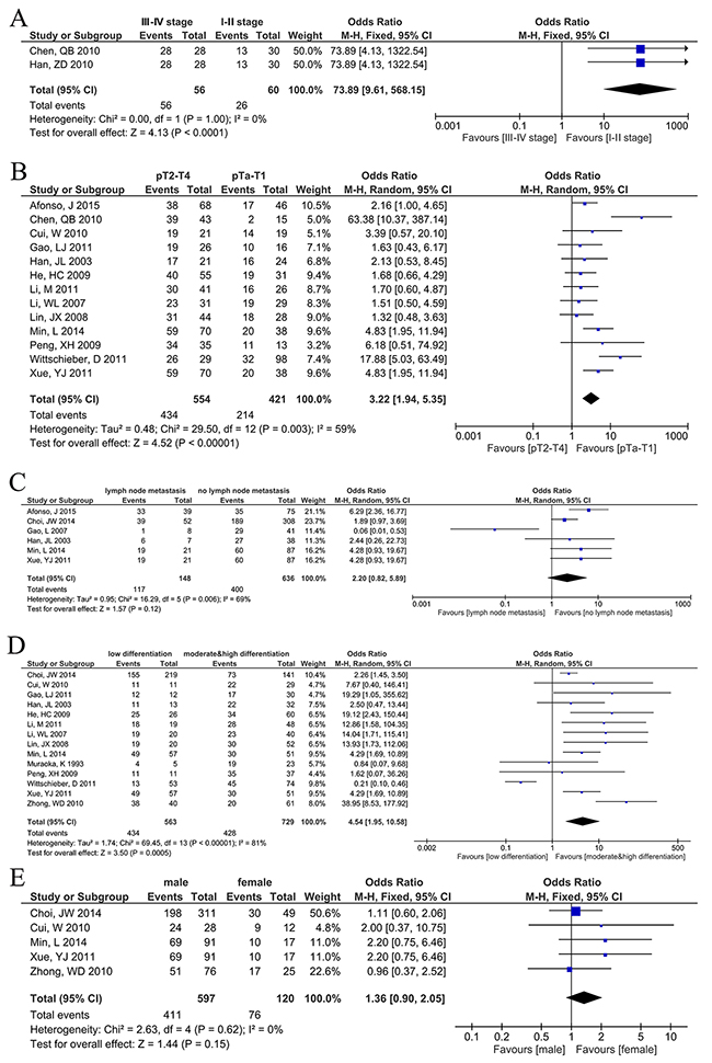
Figure 4: Forest plots of CD147 expression and clinicopathological features of bladder cancer patients. The squares and horizontal lines correspond to the study-specific OR and 95% CI. The area of the squares reflects the study-specific weight (inverse of the variance). The diamonds represent the pooled OR and 95% CI. The solid vertical line is at the null value (OR=1). (A) The relationship between CD147 expression and clinical stage. Significant difference was found between TNM III-IV stage and TNM I-II stage (OR=73.89, 95%CI=[9.61, 568.15], P<0.0001). (B) The relationship between CD147 expression and invasive depth. Significant difference was found between pT2-T4 group and pTa-T1 group (OR=3.22, 95%CI=[1.94, 5.35], P<0.00001). (C) The relationship between CD147 expression and lymph node metastasis. CD147 expression wasn’t associated with lymph node metastasis (OR=2.20, 95%CI=[0.82, 5.89], P=0.12). (D) The relationship between CD147 expression and histological differentiation. CD147 positive expression was associated with low differentiation (OR=4.54, 95%CI=[1.95, 10.58], P=0.0005). (E) The relationship between CD147 expression and gender. CD147 expression wasn’t associated with gender (OR=1.36, 95%CI=[0.90, 2.05], P=0.15).
Correlation of CD147 with invasive depth
Invasive depth also named tumor stage, from pTa to pT4, indicates deeper infiltration of the bladder wall. The association between CD147 and invasive depth was investigated in 13 studies [15, 16, 21-24, 27-31, 34, 35]. With significant heterogeneity (P=0.003, I2=59%), the random-effects model showed a significant difference between the pT2-T4 group and the pTa-T1 group (OR=3.22, 95% CI=[1.94, 5.35], P<0.00001) (Figure 4B).
CD147 expression and lymph node metastasis
Six studies [7, 15, 16, 22, 32, 35] reported the relationship between CD147 expression and lymph node metastasis of bladder cancer. Heterogeneity was observed in the analysis (P=0.006, I2=69%), and therefore, a random-effects model was used. The results showed no association between CD147 expression and lymph node metastasis (OR=2.20, 95% CI=[0.82, 5.89], P=0.12) (Figure 4C).
CD147 and histological differentiation
Histologically, bladder cancer is categorized into low, moderate, and high differentiations. Fourteen studies [7, 15, 18, 21-24, 27, 29-31, 34-36] investigated the association between CD147 expression and histological differentiation. With significant heterogeneity (P<0.00001, I2=81%), the results showed that CD147 expression was associated with low differentiation (OR=4.54, 95% CI=[1.95, 10.58], P=0.0005) (Figure 4D).
CD147 and sex
Five studies [7, 15, 18, 22, 29] involving 717 patients, reported the relationship between CD147 expression and sex. Fixed-effects model showed no difference between male and female groups (OR=1.36, 95% CI=[0.90, 2.05], P=0.15), with no heterogeneity (P=0.62, I2=0%) (Figure 4E).
Sensitivity analysis and publication bias
A sensitivity analysis was performed to evaluate the reliability of the results. While multiple datasets were available, sensitivity was tested by excluding studies one by one. Except in the case of Lymph node metastasis vs. No lymph node metastasis, all analyses were stable. To test for publication bias, we use Egger's test. All P values were higher than 0.05, indicating no publication bias. In summary, the results were stable and reliable (Table 4, Figure 5 and Figure 6).
Table 4: Summary of sensitivity and publication bias analysis.
OR/HR fluctuation |
95%CI/HR fluctuation |
Publication bias (P value) |
|
|---|---|---|---|
CD147 expression and survival analysis |
|||
OS (univariate analysis) |
2.42~3.18 |
1.74~4.45 |
0.250 |
OS (multivariate analysis) |
1.74~2.51 |
1.18~4.70 |
0.152 |
DSS (univariate analysis) |
1.54~2.05 |
1.05~3.04 |
0.205 |
DSS (multivariate analysis) |
1.43~3.04 |
0.88~6.31 |
/ |
DFS (univariate analysis) |
2.68~2.98 |
1.29~7.85 |
/ |
Recurrence vs. No recurrence |
1.79~3.32 |
1.21~7.03 |
0.465 |
CD147 expression in different bladder tissues |
|||
Bladder cancer vs. Noncancer tissues |
40.35~51.79 |
20.81~102.95 |
0.565 |
Bladder cancer vs. bladder embryonic tissues |
25~73.51 |
1.34~1297.91 |
/ |
Bladder cancer vs. Normal bladder tissues |
34.71~49.99 |
14.50~127.36 |
0.023 |
Bladder cancer vs. Para-carcinoma tissues |
52.61~68.06 |
8.39~552.26 |
/ |
CD147 expression with clinicopathological parameters |
|||
III~IV stage vs. I~II stage |
73.89~73.89 |
4.13~1322.54 |
/ |
pT2-T4 vs. pTa-T1 |
2.71~3.52 |
1.73~6.01 |
0.199 |
Lymph node metastasis vs. No lymph node metastasis |
1.65~3.13 |
0.53~8.36 |
0.653 |
Low differentiation vs. Moderate & high differentiation |
3.68~5.78 |
1.62~14.84 |
0.193 |
Male vs. Female |
1.25~1.61 |
0.80~2.82 |
0.225 |
P < 0.05, exist Publication Bias.
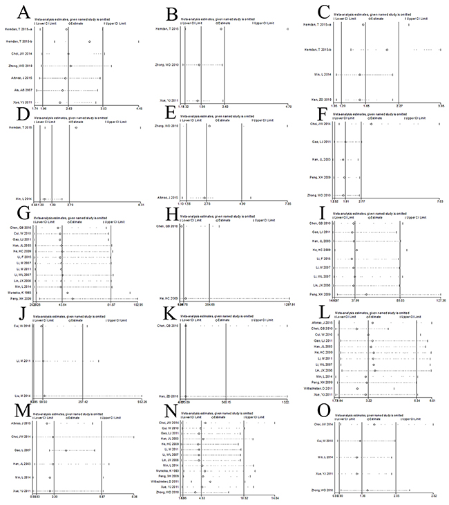
Figure 5: Sensitivity analysis. We exclude study one by one to evaluate the influences of individual studies on the final effect and all the results were consist with the result of including all studies, which means our results are stable and reliable. (A) CD147 expression and overall survival with univariate analysis; (B) CD147 expression and overall survival with multivariate analysis; (C) CD147 expression and disease specific survival with univariate analysis; (D) CD147 expression and disease specific survival with multivariate analysis; (E) CD147 expression and disease recurrence-free survival with univariate analysis; (F) CD147 expression and recurrence; (G) CD147 positive expression between bladder cancer tissues and non-cancer bladder tissues; (H) CD147 positive expression between bladder cancer tissues and bladder embryonic tissues; (I) CD147 positive expression between bladder cancer tissues and normal bladder tissues; (J) CD147 positive expression between bladder cancer tissues and para-carcinoma tissues; (K) CD147 expression and clinical stage; (L) CD147 expression and invasive depth; (M) CD147 expression and lymph node metastasis; (N) CD147 expression and histological differentiation; (O) CD147 expression and gender.
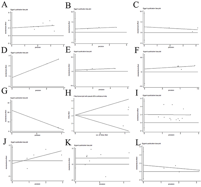
Figure 6: Publication bias. No publication bias was found by egger’s test except for CD147 positive expression between bladder cancer tissues and normal bladder tissues. (A) CD147 expression and overall survival with univariate analysis; (B) CD147 expression and overall survival with multivariate analysis; (C) CD147 expression and disease specific survival with univariate analysis; (D) CD147 expression and disease specific survival with multivariate analysis; (E) CD147 expression and recurrence; (F) CD147 positive expression between bladder cancer tissues and non-cancer bladder tissues; (G) CD147 positive expression between bladder cancer tissues and normal bladder tissues; (H) CD147 positive expression between bladder cancer tissues and normal bladder tissues (trim and fill method); (I) CD147 expression and invasive depth; (J) CD147 expression and lymph node metastasis; (K): CD147 expression and histological differentiation; (L) CD147 expression and gender.
DISCUSSION
CD147, also known as EMMPRIN, is a transmembrane glycoprotein in the immunoglobulin superfamily. It is highly expressed in many types of malignant tumors, including breast carcinoma, gastrointestinal cancer, prostate cancer, and bladder cancer, and plays a significant role in tumor proliferation, invasion, metastasis, as well as in many other processes in tumorigenesis and tumor development [37–39]. In the current meta-analysis, we pooled 25 datasets from 24 studies [4, 7, 15-24, 25-36] and demonstrated a notable association between CD147 expression and patients with bladder cancer.
According to our results, CD147 expression is closely associated with prognostic and clinicopathological characteristics of bladder cancer. Irrespective of whether the parameter tested was OS, DSS, or DFS, univariate analysis showed that patients with higher CD147 positive expression were more likely to have worse prognosis. Multivariate analysis for OS and DFS gave the same result. However, regarding multivariate analysis for DSS, our results showed no difference between CD147 positive and negative expression groups. Besides this, our results also indicated that CD147 positive expression is correlated with higher rates of recurrence. Our findings relating to survival rate were in accordance with those of other studies comparing CD147 positive and negative expression with OS, DSS, or DFS in breast, ovarian, and gastrointestinal cancers [40–42]. Results of subgroup analysis based on IHC and cut-off values were in accordance with our main results.
Furthermore, CD147 expression is associated with the response to chemotherapy for bladder cancer, and Misra et al. [43] demonstrated that CD147 enhances tumor growth and chemoresistance via the phosphatidylinositol 3-kinase (PI3K)/Akt pathway in a hyaluronan-dependent manner. Als et al. [4] found that the response rates in patients with CD147-negative and CD147-positive tumors were 74% and 39% respectively, with an odds ratio of 4.41 (95% CI=[1.91-10.1]). In addition, by silencing CD147 via RNA interference In vitro, Afonso et al. [16] showed a lower cisplatin IC50 for bladder cancer (from 24.11 μg/ml to 7.42 μg/ml). That is, the downregulation of CD147 sensitizes bladder cancer cells to chemotherapy.
As CD147 can promote the activity and expression of MCT-1 and MCT-4, forming complexes on the membrane to transport lactic acid produced by anaerobic tumor glycolysis, its expression may be increased in tumor tissues [42, 44]. As such, we investigated CD147 expression in different bladder tissues. The results showed that CD147 expression in bladder cancer tissues is greater than that in non-cancer tissues including embryonic bladder tissues, normal bladder tissues, and para-carcinoma tissues.
Next, we compared the expression of CD147 in relation to recurrence, clinical stages, invasive depth, lymph node metastasis, histological differentiation, and sex, to explore the correlation of CD147 expression with these clinicopathological parameters. In summary, CD147 expression is strongly associated with more advanced clinical stages, greater invasive depth, and poorer histological differentiation. However, CD147 expression is not correlated with lymph node metastasis or sex. As CD147 can facilitate the secretion of matrix metalloproteinase (MMP)-1, MMP-3, MMP-9, and membrane type-1 MMP, it may bring about the degradation of the basement membrane and extracellular matrix. This is one of the major mechanisms of its promotion of tumor metastasis [37, 45]. The meta-analyses of Huang et al. [41] and Peng et al. [46] also indicated that CD147 expression is associated with lymph node metastasis in gastrointestinal and prostate cancers. However, we found no association between CD147 positive expression and lymph node metastasis. However, when we excluded the study of Gao et al. [32], this result was reversed. Therefore, further qualified studies are needed to assess the relationship between CD147 and lymph node metastasis in bladder cancer.
Besides the immunohistochemistry of CD147 protein levels analyzed in this study, other researches are available. Using Western blotting, Xue et al. [15] showed higher CD147 expression in bladder cancer cell lines T24, SCaBER, 5637, BIU-87, and J82 than in the normal urothelial cell line SV-HUC-1. In particular, the T24 and SCaBER cell lines had the highest CD147 protein levels. Bhagirath et al. [26] compared the serum concentration of CD147 in bladder cancer patients and healthy people by enzyme-linked immunosorbent assay and found a significant increase in CD147 levels in the serum samples of bladder cancer patients. Similarly, Li et al. [21] demonstrated higher CD147 mRNA levels in bladder transitional cell carcinoma (59.7%) than in normal bladder tissue (0%), but found no difference with clinical stage (r=0.048, P=0.698) or histological differentiation (r=0.222, P=0.071). Min et al. [22] also found upregulation of CD147 in bladder cancer at both mRNA and protein levels (mRNA: bladder cancer tissues 0.967±0.133, paracarcinoma tissues 0.223±0.096; protein: bladder cancer tissues 0.766±0.103, paracarcinoma tissues 0.165±0.055).
To our knowledge, this meta-analysis is the first to report the relationship between CD147 and bladder cancer. We analyzed 24 high-quality studies (NOS≥7 points) with significant results. Meanwhile, some limitations need to be acknowledged. First, our analysis is based upon published studies found in the literature, but we failed to obtain any unpublished data. Second, patients we included are mostly from China, which limits the universality of our results. Third, different studies used different criteria for CD147 expression, with different follow-up times. This may result in some bias. Nevertheless, we conducted subgroup analyses based on IHC and cut-off values, and this indicates that our results were reliable.
In this meta-analysis, we demonstrated that CD147 expression is increased in bladder cancer tissues compared with non-cancer tissues. This is strongly correlated with poorer OS, DSS, and DFS; recurrence; advanced clinical stages; greater invasive depth; and poorer histological differentiation. However, it was not correlated with lymph node metastasis or sex. In summary, CD147 could be an important diagnostic and prognostic biomarker for bladder cancer.
MATERIALS AND METHODS
Search strategy
We searched PubMed (1966-2016), EMBASE (1980-2016), the Cochrane Library (1996-2016), Web of Science (1945-2016), China National Knowledge Infrastructure (1982-2016), and the WanFang databases (1988-2016). The studies were restricted to humans, but not restricted by date, language, or publication status. The following combined search terms were used: (Bladder Neoplasm*, Bladder Tumor*, Bladder Cancer*, Bladder Carcinoma, Bladder Transitional Cell Carcinoma, Bladder Squamous Cell Carcinoma, Bladder Adenocarcinoma) AND (CD147, Extracellular matrix metalloproteinase inducer, EMMPRIN, BSG). We combined the term appropriately with MeSH Terms and used an appropriate adjustment for different databases. Details of the search strategies can be found in Appendix 1.
Criteria for including studies
1. Published or unpublished case control study or cohort study in English or Chinese with the full text available;
2. All cases had survival data or clinical pathological characteristic data, without radiotherapy or chemotherapy or biological therapy before sampling;
3. Diagnosis of bladder cancer was proven by pathological methods;
4. Studies of CD147 expression based on primary bladder cancer tissue (via either biopsy or surgical), rather than serum or any other kinds of indirect specimen were included;
5. The best quality study was retained for dealing duplicated study.
Criteria for excluding studies
1. Cell or animal studies, case reports, letters, reviews;
2. The standard of pathological diagnosis was not clear.
Assessment of included studies
The Newcastle-Ottawa quality assessment scale of case control studies (NOS) [47] was adopted to assess the quality of included studies. It has three categories (selection, comparability, and exposure) and eight items. The quality assessment values ranged from 0 to 9 stars. Studies scored more than 6 stars was included for our analysis.
Statistical analysis
Literature were independently filtered by two authors to exclude unrelated studies. Then, full texts were independently reviewed, and controversies were solved by discussion. Data were extracted independently by two authors. The software Revman 5.3 and Stata 14.0 were applied. Results were showed with odds ratios (OR), standard mean difference (SMD) or HR (hazard ratio) with 95% confidence intervals (95% CI). Fixed-effects model was adopted for non-heterogeneous data (P > 0.1 and I2 < 50%); otherwise, random-effects model was used. If possible, heterogeneity was explored and subgroup analyses were performed.
Additionally, sensitivity analysis was performed to evaluate the influences of individual studies on the final effects size if the parameter has no less than three datasets for parameter. Otherwise, analysis based on different models was used.
Finally, publication bias was assessed by Egger’s test (P < 0.05 was considered statistically significant). If publication bias was confirmed, a trim-and-fill method developed by Duval and Tweedie was implemented to adjust for this bias. Then, we replicated the funnel plot with their ‘‘missing’’ counterparts around the adjusted summary estimates [48].
Abbreviations
OR: odds ratio
SMD: standard mean difference
HR: hazard ratio
MCT: mono-carboxylate transporter
EMMPRIN: extracellular matrix metalloproteinase inducer
DSS: disease specific survival
IHC: immunohistochemistry
NOS: Newcastle-Ottawa quality assessment scale
OS: overall survival
CI: confidence intervals
DFS: disease recurrence-free survival
SP: streptavidin-perosidase
PI3K: phosphatidylinositol 3-kinase
IC: inhibitory concentration
MMP: matrix metalloproteinase
BSG: basigin
Author contributions
Hongru Li did literature searching, data extraction, quality assessment, statistical analysis and drafted the paper. Yadong Xu participated in literature searching, data extraction, quality assessment and drafting the paper. Hui Li conceived the study and participated in data extraction and statistical analysis.
CONFLICTS OF INTEREST
All authors disclosed no relevant relationships.
FUNDING
No funding.
REFERENCES
1. May M. Statistics: attacking an epidemic. Nature. 2014;509:S50-S51.
2. Siegel RL, Miller KD, Jemal A. Cancer statistics, 2016. CA Cancer J Clin. 2016;66:7-30.
3. Kaufman DS, Shipley WU, Feldman AS. Bladder cancer. Lancet. 2009;374:239-249.
4. Als AB, Dyrskjot L, von der Maase H, Koed K, Mansilla F, Toldbod HE, Jensen JL, Ulhoi BP, Sengelov L, Jensen KM, Orntoft TF. Emmprin and survivin predict response and survival following cisplatin-containing chemotherapy in patients with advanced bladder cancer. Clin Cancer Res. 2007;13:4407-4414.
5. Shariat SF, Ashfaq R, Karakiewicz PI, Saeedi O, Sagalowsky AI, Lotan Y. Survivin expression is associated with bladder cancer presence, stage, progression, and mortality. Cancer. 2007;109:1106-1113.
6. Bi J, Chen X, Zhang Y, Li B, Sun J, Shen H, Kong C. Fascin is a predictor for invasiveness and recurrence of urothelial carcinoma of bladder. Urol Oncol. 2012;30:688-694.
7. Choi JW, Kim Y, Lee JH, Kim YS. Prognostic significance of lactate/proton symporters MCT1, MCT4, and their chaperone CD147 expressions in urothelial carcinoma of the bladder. Urology. 2014;84:245-249.
8. Yan L, Zucker S, Toole BP. Roles of the multifunctional glycoprotein, emmprin (basigin; CD147), in tumour progression. Thromb Haemost. 2005;93:199-204.
9. Caudroy S, Polette M, Tournier JM, Burlet H, Toole B, Zucker S, Birembaut P. Expression of the extracellular matrix metalloproteinase inducer (EMMPRIN) and the matrix metalloproteinase-2 in bronchopulmonary and breast lesions. J Histochem Cytochem. 1999;47:1575-1580.
10. Gou X, Chen H, Jin F, Wu W, Li Y, Long J, Gong X, Luo M, Bi T, Li Z, He Q. Expressions of CD147, MMP-2 and MMP-9 in laryngeal carcinoma and its correlation with poor prognosis. Pathol Oncol Res. 2014;20:475-481.
11. Ishibashi Y, Matsumoto T, Niwa M, Suzuki Y, Omura N, Hanyu N, Nakada K, Yanaga K, Yamada K, Ohkawa K, Kawakami M, Urashima M. CD147 and matrix metalloproteinase-2 protein expression as significant prognostic factors in esophageal squamous cell carcinoma. Cancer. 2004;101:1994-2000.
12. Kim Y, Choi JW, Lee JH, Kim YS. Expression of lactate/H(+) symporters MCT1 and MCT4 and their chaperone CD147 predicts tumor progression in clear cell renal cell carcinoma: immunohistochemical and the Cancer Genome Atlas data analyses. Hum Pathol. 2015;46:104-112.
13. Yu YH, Morales J, Feng L, Lee JJ, El-Naggar AK, Vigneswaran N. CD147 and Ki-67 overexpression confers poor prognosis in squamous cell carcinoma of oral tongue: a tissue microarray study. Oral Surg Oral Med Oral Pathol Oral Radiol. 2015;119:553-565.
14. Zeng HZ, Qu YQ, Liang AB, Deng AM, Zhang WJ, Xiu B, Wang H, Wang H. Expression of CD147 in advanced non-small cell lung cancer correlated with cisplatin-based chemotherapy resistance. Neoplasma. 2011;58:449-454.
15. Xue YJ, Lu Q, Sun ZX. CD147 overexpression is a prognostic factor and a potential therapeutic target in bladder cancer. Med Oncol. 2011;28:1363-1372.
16. Afonso J, Santos LL, Miranda-Goncalves V, Morais A, Amaro T, Longatto-Filho A, Baltazar F. CD147 and MCT1-potential partners in bladder cancer aggressiveness and cisplatin resistance. Mol Carcinog. 2015;54:1451-1466.
17. Hemdan T, Malmstrom PU, Jahnson S, Segersten U. Emmprin expression predicts response and survival following cisplatin containing chemotherapy for bladder cancer: a validation study. J Urol. 2015;194:1575-1581.
18. Zhong WD, Chen QB, Ye YK, Han ZD, Bi XC, Dai QS, Liang YX, Zeng GH, Wang YS, Zhu G, Chen ZN, He HC. Extracellular matrix metalloproteinase inducer expression has an impact on survival in human bladder cancer. Cancer Epidemiol. 2010;34:478-482.
19. Han ZD, He HC, Bi XC, Qin WJ, Dai QS, Zou J, Ye YK, Liang YX, Zeng GH, Zhu G, Chen ZN, Zhong WD. Expression and clinical significance of CD147 in genitourinary carcinomas. J Surg Res. 2010;160:260-267.
20. El-Rehim DM, El-Maqsoud NM, El-Hamid AM, El-Bab TK, Galal EM. Expression of extracellular matrix metalloproteinase inducer and fascin in urinary bladder cancer: correlation with clinicopathological characteristics. Mol Clin Oncol. 2013;1:297-304.
21. Li M, Xie Q, Yang QT, Lin BS, Zhang C, Zheng JH. The Expression and clinical significance of CD147mRNA and protein in human bladder transitional cell carcinoma. J Clin Urol. 2011:433-436.
22. Min L, Mao XM, Xue YJ, Zou XF, Yuan YH, Xiao RH, Zhang GX, Wu GQ, Wang XN. Expression and clinical significance of extracellular matrix metalloproteinase inducer molecule CD147 in bladder urothelial carcinoma tissue. Chongqing Med. 2014:4574-4578.
23. Peng XH, Zu XB, Liang PY, Ou SJ. Expression of MMP-2, MMP-9 and CD147 and their relationships in bladder transitional cell carcinoma. J Hainan Med Coll. 2009:841-844.
24. Gao LJ. Expression and clinical significance of CD147 and MDM2 in bladder tumor. Tianjin Med U. 2011:51.
25. Li F, Ye XD, Lian XX, Feng YP, Liao YJ. Expression and correlation of CD147 protein and apoptosis inhibiting gene Survivin in bladder cancer. J Contempr Urol Reprod Oncol. 2015;1:44-46.
26. Bhagirath D, Abrol N, Khan R, Sharma M, Seth A, Sharma A. Expression of CD147, BIGH3 and Stathmin and their potential role as diagnostic marker in patients with urothelial carcinoma of the bladder. Clin Chim Acta. 2012;413:1641-1646.
27. Wittschieber D, Stenzinger A, Klauschen F, Stephan C, Jung K, Erbersdobler A, Rabien A. Decreased RECK and Increased EMMPRIN expression in urothelial carcinoma of the bladder are associated with tumor aggressiveness. Pathobiology. 2011;78:123-131.
28. Chen QB, Bi XC, He HC, Han ZD, Ye YK, Liang YX, Wang YS, Zeng GH, Zhong WD. Relationship between the expression of CD147 and clinic pathological data in bladder cancer. Chin J Clin. 2010;4:273-277.
29. Cui W. The expression and clinical significance of EMMPRIN and MMP-9 in urothelial carcinoma of the bladder. ZunYi Medical University. 2010:34.
30. He HC. Relationship between the expression of CD147 and clinic pathological data in bladder cancer. Guangzhou Medical College. 2009:38.
31. Lin JX, Li M, Lin BS, Zhang C. The expression and clinical significance of CD147, MMP-2 in bladder transitional cell carcinoma. Chin J Coal Ind Med. 2008;6:808-809.
32. Gao L, Wang XM, Lin XG, Wang XW. Expression and significance of matrix metalloproteinase-2 and CD147 in transitional cell carcinoma of bladder. Chin J Lab Diag. 2007;9:1225-1227.
33. Li M, Zhang ZB, Lin BS, Zhuang RH, Zhang C, Zheng JH. The expression and clinical significance of CD147, MMP-2 and VEGF in human bladder transitional cell carcinoma. Chin J Exp Surg. 2007;4:487-488.
34. Li WL, Duan JM, Liu DJ, Ma BL, Lu JZ, Zuo LJ, Qin DS. The expression and clinical significance of EMMPRIN and MMP-7 in bladder transitional cell carcinoma. Shandong Med J. 2007:39-40.
35. Han JL, Xie WL, Huang J. Expression and clinical significance of matrix metalloproteinase-2 and CD147 in bladder transitional cell carcinoma. Chin J Exp Surg. 2003;12:1066-1067.
36. Muraoka K, Nabeshima K, Murayama T, Biswas C, Koono M. Enhanced expression of a tumor-cell-derived collagenase-stimulatory factor in urothelial carcinoma: Its usefulness as a tumor marker for bladder cancers. Int J Cancer. 1993;55:19-26.
37. Grass GD, Toole BP. How, with whom and when: an overview of CD147-mediated regulatory networks influencing matrix metalloproteinase activity. Biosci Rep. 2015;36:e283.
38. Li H, Wu D, Shi S, Xu Y, Wei L, Liu J, Liu Y. Expression and clinical significance of CD147 in renal cell carcinoma: a meta-analysis. Oncotarget. 2017;8:51331-51344. https://doi.org/10.18632/oncotarget.17376.
39. Li H, Xi Z, Dai X, Wu W, Li Y, Liu Y, Zhang H. CD147 and glioma: a meta-analysis. J Neurooncol. 2017.
40. Bovenzi CD, Hamilton J, Tassone P, Johnson J, Cognetti DM, Luginbuhl A, Keane WM, Zhan T, Tuluc M, Bar-Ad V, Martinez-Outschoorn U, Curry JM. Prognostic indications of elevated MCT4 and CD147 across cancer types: a Meta-Analysis. Biomed Res Int. 2015;2015:242437.
41. Huang X, Shen W, Xi H, Zhang K, Cui J, Wei B, Chen L. Prognostic role of extracellular matrix metalloproteinase inducer/CD147 in gastrointestinal cancer: a meta-analysis of related studies. Oncotarget. 2016;7:81003-81011. https://doi.org/10.18632/oncotarget.12745.
42. Xin X, Zeng X, Gu H, Li M, Tan H, Jin Z, Hua T, Shi R, Wang H. CD147/EMMPRIN overexpression and prognosis in cancer: a systematic review and meta-analysis. Sci Rep. 2016;6:32804.
43. Misra S, Ghatak S, Zoltan-Jones A, Toole BP. Regulation of multidrug resistance in cancer cells by hyaluronan. J Biol Chem. 2003; 278:25285-25288.
44. Li S, Nguyen TT, Bonanno JA. CD147 required for corneal endothelial lactate transport. Invest Ophthalmol Vis Sci 2014;55:4673-4681.
45. Huang P, Chang S, Jiang X, Su J, Dong C, Liu X, Yuan Z, Zhang Z, Liao H. RNA interference targeting CD147 inhibits the proliferation, invasiveness, and metastatic activity of thyroid carcinoma cells by down-regulating glycolysis. Int J Clin Exp Pathol. 2015;8:309-318.
46. Peng F, Li H, Ning Z, Yang Z, Li H, Wang Y, Chen F, Wu Y. CD147 and prostate cancer: a systematic review and meta-analysis. PLos One. 2016;11:e163678.
47. Wells GA, Shea B, O' Connell D, Peterson JEA, Welch V, Losos M, Tugwell P. The Newcastle-Ottawa Scale (NOS) for assessing the quality of nonrandomised studies in meta-analyses. 2000.
48. Duval S, Tweedie R. Trim and fill: a simple funnel-plot-based method of testing and adjusting for publication bias in meta-analysis. Biometrics. 2000;56:455-463.