
INTRODUCTION
Long non-coding RNAs (lncRNAs) are a group of RNA transcripts that exceed 200 nt in length yet lack significant open reading frames (ORFs) [1]. They regulate gene expression through transcriptional, post-transcriptional and epigenetic effects [2-8]. Tens of thousands of lncRNAs have been identified in the human genome [9], many of which are abnormally expressed in a variety of human tumors, and are involved in various stages of carcinogenesis, including tumor initiation, progression and metastasis [10-17]. However, the function of the vast majority of these lncRNAs is still unclear.
In a previous study, we performed gene expression profile (GEP) analysis by microarray and found that one lncRNA named actin filament-associated protein 1 antisense RNA1 (AFAP1-AS1) was significantly upregulated in nasopharyngeal carcinoma (NPC), and promoted invasion and metastasis of cancer cells by regulating the expression of several small GTPase family members and molecules in the actin cytokeratin signaling pathway [18]. However, it is not yet known whether there is any other biological function of AFAP1-AS1 in the tumorigenesis of NPC.
In this study, using GEP dataset, we found that a key molecular maker of tumor immune evasion, programmed death 1 (PD-1), was positively correlated with the expression of AFAP1-AS1. Therefore, we used in situ hybridization to detect the expression of AFAP1-AS1 and immunohistochemical staining to detect the expression of PD-1 in a cohort of 96 NPC biopsies, and we analyzed co-expression of AFAP1-AS1 and PD-1 and its relevance in clinical outcomes and prognosis. The results suggest that AFAP1-AS1 might be involved in the PD-1 immune checkpoint pathway and that PD-1 and AFAP1-AS1 might jointly promote the formation and development of NPC.
RESULTS
The expression of AFAP1-AS1 is positively correlated with PD-1 in NPC
The Gene Expression Omnibus (GEO) database [19] is a public gene expression data repository that serves as a valuable data repository for biomedical research and has collected a large amount of gene expression data for data mining [20]. Mining of published high-throughput data is a commonly used and low-cost method for identifying novel biomarkers and gaining insight into the biological functions of novel genes [21-23]. To identify potential novel functions of AFAP1-AS1, we downloaded a GEP dataset, GSE12452, from the GEO database; this dataset consists of 41 samples of whole-genome GEP data, including 10 samples of non-tumor nasopharyngeal epithelial (NPE) biopsies and 31 cases of NPC [24]. We found that there were 4196 differentially expressed genes in the GSE12452 dataset. Among these differentially expressed genes, AFAP1-AS1 was highly expressed in NPC cells and was positively correlated with the expression of PD-1, a key molecular marker of tumor immune evasion (Figure 1, P=0.05).
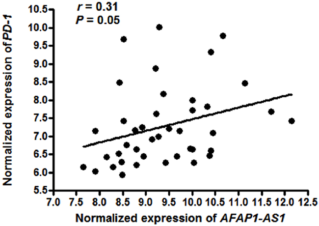
Figure 1: Expression of AFAP1-AS1 is positively correlated with PD-1. Normalized gene expression signatures of AFAP1-AS1 and PD-1 were derived from whole-genome GEP dataset GSE12452. Expression of AFAP1-AS1 and PD-1 was positively correlated in 31 NPC samples and 10 NPE samples (r=0.31, P=0.05).
AFAP1-AS1 and PD-1 are co-expressed in infiltrating lymphocytes in NPC tissue
Since PD-1 is a membrane protein and mainly expressed on the lymphocyte cell surface [25]; and tumor-infiltrating lymphocytes are associated with the development and progression of NPC [26]. we set out to assess AFAP1-AS1 and PD-1 expression in a cohort of 96 paraffin-embedded NPC samples via in situ hybridization and immunohistochemical staining, respectively (Supplementary Table 1). Expression of AFAP1-AS1 was absent or very low in adjacent non-tumor NPE (Figure 2A) but high in NPC cells and infiltrating lymphocytes in 68 of 96 cases (70.8%, Figure 2B). Similarly, the expression of PD-1 was low or negative in non-tumor NPE (Figure 2C) but high in infiltrating lymphocytes surrounding NPC cells (36 of 96 cases, 37.5%, Figure 2D).
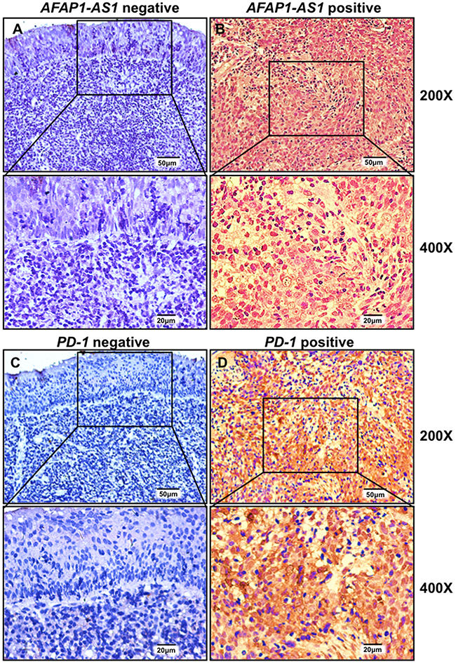
Figure 2: AFAP1-AS1 and PD-1 are highly and jointly expressed in infiltrating lymphocytes of NPC tissues. Representative images of in situ hybridization for AFAP1-AS1 (A & B) and immunohistochemical staining for PD-1 (C & D) are shown. AFAP1-AS1 and PD-1 are negatively expressed in adjacent non-tumor NPE tissue (A & C) but highly expressed in infiltrating lymphocytes (B & D).
High expression of AFAP1-AS1 or PD-1 is correlated with distant metastasis at relapse
We then analyzed the correlation between the expression of AFAP1-AS1 and PD-1 and clinicopathological features of these 96 NPC patients. There was no significant correlation between the expression of AFAP1-AS1 or PD-1 and patients’ gender, age at diagnosis, tumor size (T stage), lymphatic invasion (N stage), distant metastasis at diagnosis (M stage) and overall clinical staging (Supplementary Table 2), but patients with high expression of AFAP1-AS1 or PD-1 in NPC-infiltrating lymphocytes were more likely to have distant metastasis when they relapsed (Figure 3A and 3B, P=0.005 and P=0.020, respectively).
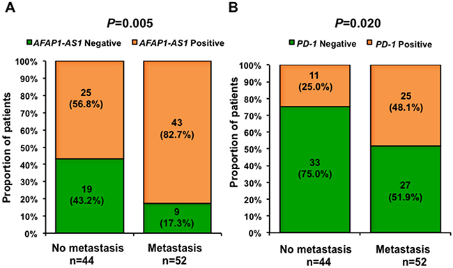
Figure 3: Expression of AFAP1-AS1 or PD-1 is associated with distant metastasis. Patients with high expression of AFAP1-AS1 (A) or PD-1 (B) in NPC tumor-infiltrating lymphocytes were more likely to have distant metastasis when they relapsed.
Co-expression of AFAP1-AS1 and PD-1 predicts poor prognosis of NPC
Finally, we analyzed the association of AFAP1-AS1 and PD-1 expression with NPC patients’ outcomes. Patients with positive expression of AFAP1-AS1 or PD-1 in NPC biopsies had a poor prognosis, with shorter overall survival (47.7% five-year survival with positive expression of AFAP1-AS1 vs. 92.8% with negative expression, P=0.001, Figure 4A; 52.8% five-year survival with positive PD-1 expression vs. 65.0% with negative expression, P=0.049, Figure 4B). NPC patients with positive expression of both AFAP1-AS1 and PD-1 had much shorter overall survival (38.5% five-year survival, P=0.002, Figure 5).
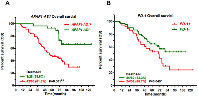
Figure 4: High expression of AFAP1-AS1 or PD-1 predicts poor prognosis. Kaplan-Meier survival curves of patients with NPC, stratified by AFAP1-AS1 expression (A) and PD-1 expression (B), shows that high expression of AFAP1-AS1 or PD-1 predicts poor prognosis.
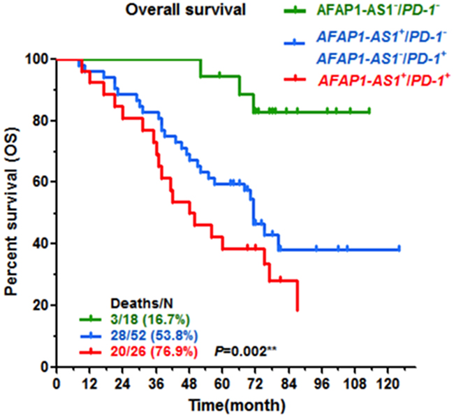
Figure 5: Co-expression of AFAP1-AS1 and PD-1 predicts the poorest outcomes. Patients with high expression of both AFAP1-AS1 and PD-1 had a significantly less favorable prognosis than those with low expression of AFAP1-AS1 and/or PD-1.
DISCUSSION
NPC is an Epstein-Barr Virus (EBV)-associated malignancy and is the most common malignant head and neck tumor, originating in the nasopharyngeal epithelium [27-31]. High incidences of NPC are observed in Southeast Asia and southern China, resulting in serious healthcare problems in these regions [32-34]. NPC is highly metastatic and heterogeneous compared with other head and neck tumors [35-40]. Radiotherapy has been used as the primary clinical treatment for all stages of NPC over the past several decades, but many patients eventually die due to recurrence and distant metastasis [41-43]. Although recent studies have shown that induction chemotherapy plus concurrent chemoradiotherapy significantly improves failure-free survival in locally or regionally advanced NPC with acceptable toxicity, long-term efficacy and toxicities remain unclear [44]. Management of advanced NPC is therefore a highly challenging issue, and novel and effective therapies for NPC are urgently needed.
Recently, tumor immune evasion has emerged as a hallmark of cancer progression [45-47]. Immune surveillance is an important mechanism in preventing the development of cancer and inhibiting tumor growth and metastasis [48-50]. There are many immunosuppressive mechanisms in the tumor microenvironment that can decrease the activity of tumor-infiltrating lymphocytes and increase the risk of tumor metastasis and recurrence [51]. Among these mechanisms, T cell-mediated immune responses, especially CD8+ cytotoxic T lymphocytes, play an important role in tumor immunity [52, 53].
PD-1 is a transmembrane receptor that is mainly expressed on T cells. It was first cloned in T cell hybridomas and was named “programmed death receptor” because of its involvement in T cell apoptosis [25]. In tumor tissues, PD-1 is mainly expressed in tumor-infiltrating lymphocytes (TILs) [54, 55]. High expression of PD-1 in TILs led to depletion and deactivation of T cells [56-60]. PD-1 also interacts with programmed death ligand-1 (PD-L1) and programmed death ligand-2 (PD-L2), which are mainly expressed on the surface of tumor cells or in the tumor matrix [61, 62]; these ligands activate PD-1, which then inhibits the proliferation of T cells and promotes the immune escape of tumor cells, playing an important role in immune suppression and cancer progression [63-65]. The blockade of immune checkpoints has been the most promising approach to activating antitumor immunity. A tumor immunotherapy treatment strategy using a combined PD-1/PD-L1 antibody has entered the stage of clinical trials and shown good performance [66, 67].
It has been reported that local infiltration of T cells is a favorable indicator of survival in NPC patients, but many studies have indicated that NPC can escape immune surveillance through various mechanisms [68-75]. Recent studies have shown that NPC has high levels of PD-L1 and PD-1, indicating that NPC may be a candidate for PD-1/PD-L1-dericted therapies [76-78]. However, the underlying mechanism of PD-1 regulation in NPC is undetermined.
In a previous study, we found that the lncRNA AFAP1-AS1 is significantly upregulated in NPC and promotes invasion and metastasis of cancer cells [18]. Interestingly, using the GEO database, we found and confirmed that the expression of AFAP1-AS1 is positively correlated with PD-1, that high expression of PD-1 and AFAP1-AS1 predicts high incidence of recurrence or metastasis and that co-expression of AFAP1-AS1 and PD-1 in NPC biopsies predicted the poorest prognosis.
However, there are still several relevant mechanism-related questions to be solved urgently. For example, is there a regulatory relationship between AFAP1-AS1 and PD-1? Does AFAP1-AS1 promote the expression of PD-1? And how does AFAP1-AS1 regulate PD-1? We speculate that AFAP1-AS1 may regulate PD-1 expression through the following mechanisms. First, AFAP1-AS1 may act as a competing endogenous RNA (ceRNAs) [79-81] to regulate PD-1 expression. Second, AFAP1-AS1 may bind to certain transcriptional complexes to regulate PD-1 transcription. Third, AFAP1-AS1 may affect epigenetic modification of PD-1. These questions warrant in-depth exploration in future studies.
In conclusion, to our knowledge, this is the first study to explore the co- expression of a lncRNA, AFAP1-AS1, and an immune escape marker, PD-1, in tumor-infiltrating lymphocytes among NPC patients, as well as their synergistic effect on prognosis. This study provides two potential therapeutic targets for NPC, AFAP1-AS1 and PD-1, to inhibit tumor metastasis and stimulate anti-tumor immunity. Patients with higher expression of both AFAP1-AS1 and PD-1 might be ideal candidates for future clinical trials of anti-PD-1 therapy. Our study is limited by its retrospective nature, with a relatively small sample size. Further studies with larger sample sizes are warranted.
MATERIALS AND METHODS
Tissue samples
A total of 96 samples of paraffin-embedded NPC tissue were collected from newly diagnosed NPC patients at the Xiangya Hospital and the Affiliated Cancer Hospital of Central South University (Changsha China). All specimens were confirmed by histopathological examination. All of the patients had received routine radiotherapy. This study was approved by the Research Ethics Board of Xiangya Hospital and the Affiliated Cancer Hospital of Central South University, and signed informed consent was obtained from each participant before they were enrolled in the study. Clinicopathological data were collected from patient medical records and are reported in Supplementary Table 1.
In situ hybridization
In situ hybridization was performed to detect the expression of AFAP1-AS1 in tissue specimens using three 30-nucleotide probes from different regions of AFAP1-AS1. GAPDH was used as a positive control. The probe sequences were as follows.
AFAP1-AS1 probes:
Probe 1: 5’- ATTCCTTTATTTTATGGGATGTTCTGTAGGGAGTT-3’,
Probe 2: 5’-TAGAAATGAGAAAAGAATCACCAAGAGAGTAAGCA -3’,
Probe 3: 5’-CCCTACAGCTAGTTTCCTCTTCATTTATTCATTT-3’
GAPDH probes:
Probe 1: 5’-CCACTTTACCAGAGTTAAAAGCAGCCCTGG-3’
Probe 2: 5’-CAGTAGAGGCAGGGATGATGTTCTGGAGAG-3’
Probe 3: 5’-GTCAGAG GAGACCACCTGGTG CTCAGTGTA-3’
The probes were synthesized and labeled with DIG-dUTP at the 3’ end using a kit from Invitrogen (Shanghai, China) [82-84]. The in situ hybridization results were independently scored manually by two pathologists who counted 20 sequential high-power fields judged to be representative of the tumor, while remaining blinded to clinical information.
Immunohistochemistry
Paraffin-embedded sections (3 μm) were used for PD-1 staining. Paraffin sections were dewaxed using turpentine and gradient alcohol, immersed in 3% H2O2 at room temperature for 10 min and then treated with citric acid buffer [85, 86]. Staining for PD-1 (Proteintech, Wuhan, China) was observed under the microscope. Samples were divided into a PD-1-negative group and a PD-1-positive group by double-blind scoring by two pathologists.
Data analysis
We downloaded an NPC gene expression dataset from the GEO database (accession number GSE12452). The GSE12452 microarray consists of 10 non-tumor NPE biopsies and 31 cases of NPC [24]. We used Significant Analysis of Microarray (SAM) software [87] to analyze the microarray expression profiles(cut-off=1.5, FDR<0.05 lncRNA expression) and selected differentially expressed molecules of interest for the subsequent Pearson correlation analysis.
Pearson correlation analysis was used to evaluate the expression levels of AFAP1-AS1 and PD-1. The Chi-squared test was used to evaluate the expression of AFAP1-AS1, PD-1 and clinicopathological features such as gender, age at diagnosis, TNM staging, and metastasis, among others. Survival analysis was performed using the Kaplan-Meier test. A threshold of P<0.05 was used to indicate statistical significance, and all tested P values were two-sided. Statistical analysis was performed using SPSS 13 and GraphPad 5 software
Abbreviations
NPC: nasopharyngeal carcinoma; LncRNAs: Long noncoding RNAs; AFAP1-AS1: actin lament associated protein 1 antisense RNA1; PD-1: programmed death 1; ORF: open reading frame; GEP: gene expression profile; GEO: Gene Expression Omnibus; NPE: nasopharyngeal epithelial; EBV: Epstein-Barr Virus; TILs: tumor infiltrating lymphocytes; PD-L1: programmed death ligand-1; PD-L2: programmed death ligand-2; ceRNAs: Competing Endogenous RNAs; SAM: Significant Analysis of Microarray.
Author contributions
Z.Z. and S.L. designed the project and revised the manuscript. Y.T., W.X. and Z.Z drafted the manuscript. Y.T., Y.H., S.L., L.Y., J.W., Y.L., C.F., P.Z. C.G., S.Z., Z.G. X.L. and F.X. conducted the experiments and analyzed the data. X.L., G.L., Y.L. and W.X. participated in designing the experiments, analyzing the data and revising the manuscript. All the authors read and approved the final version of the paper.
CONFLICTS OF INTEREST
The authors declare that there are no conflicts of interest in this work.
FUNDING
This work was supported in part by the National Natural Science Foundation of China (81201523, 81372907, 81301757, 81472531, 81402009, 81572787, 81672683 and 81672993), the Natural Science Foundation of Hunan Province (14JJ1010, 2015JJ1022 and 2016JC2035), the Mittal Innovation Foundation of Central South University (15MX46 and MX2016442), and the Fundamental Research Funds for Central Universities of Central South University (2014zzts066 and 2016zzts478).
REFERENCES
1. Gong Z, Zhang S, Zhang W, Huang H, Li Q, Deng H, Ma J, Zhou M, Xiang J, Wu M, Li X, Xiong W, Li X, et al. Long non-coding RNAs in cancer. Sci China Life Sci. 2012; 55:1120–24.
2. Engreitz JM, Haines JE, Perez EM, Munson G, Chen J, Kane M, McDonel PE, Guttman M, Lander ES. Local regulation of gene expression by lncRNA promoters, transcription and splicing. Nature. 2016; 539:452–55.
3. Gong Z, Yang Q, Zeng Z, Zhang W, Li X, Zu X, Deng H, Chen P, Liao Q, Xiang B, Zhou M, Li X, Li Y, et al. An integrative transcriptomic analysis reveals p53 regulated miRNA, mRNA, and lncRNA networks in nasopharyngeal carcinoma. Tumour Biol. 2016; 37:3683–95.
4. Lian Y, Li XY, Tang YY, Yang LT, Li XL, Xiong W, Li GY, Zeng ZY. Long Non-coding RNAs Function as Competing Endogenous RNAs to Regulate Cancer Progression. Prog Biochem Biophys. 2016; 43:219–25.
5. Gong Z, Zhang S, Zeng Z, Wu H, Yang Q, Xiong F, Shi L, Yang J, Zhang W, Zhou Y, Zeng Y, Li X, Xiang B, et al. LOC401317, a p53-regulated long non-coding RNA, inhibits cell proliferation and induces apoptosis in the nasopharyngeal carcinoma cell line HNE2. PLoS One. 2014; 9:e110674.
6. Xu K, Xiong W, Zhou M, Wang H, Yang J, Li X, Chen P, Liao Q, Deng H, Li X, Li G, Zeng Z. Integrating ChIP-sequencing and digital gene expression profiling to identify BRD7 downstream genes and construct their regulating network. Mol Cell Biochem. 2016; 411:57–71.
7. Wang Y, Mo Y, Gong Z, Yang X, Yang M, Zhang S, Xiong F, Xiang B, Zhou M, Liao Q, Zhang W, Li X, Li X, et al. Circular RNAs in human cancer. Mol Cancer. 2017; 16:25.
8. Tang Y, Wang J, Lian Y, Fan C, Zhang P, Wu Y, Li X, Xiong F, Li X, Li G, Xiong W, Zeng Z. Linking long non-coding RNAs and SWI/SNF complexes to chromatin remodeling in cancer. Mol Cancer. 2017; 16:42.
9. Tang K, Wei F, Bo H, Huang HB, Zhang WL, Gong ZJ, Li XY, Song YL, Liao QJ, Peng SP, Xiang JJ, Zhou M, Ma J, et al. Cloning and Functional Characterization of a Novel Long Non-coding RNA Gene Associated With Hepatocellular Carcinoma. Prog Biochem Biophys. 2014; 41:153–62.
10. Yu J, Liu Y, Gong Z, Zhang S, Guo C, Li X, Tang Y, Yang L, He Y, Wei F, Wang Y, Liao Q, Zhang W, et al. Overexpression long non-coding RNA LINC00673 is associated with poor prognosis and promotes invasion and metastasis in tongue squamous cell carcinoma. Oncotarget. 2017; 8:16621–32. doi: 10.18632/oncotarget.14200.
11. Yang L, Tang Y, He Y, Wang Y, Lian Y, Xiong F, Shi L, Zhang S, Gong Z, Zhou Y, Liao Q, Zhou M, Li X, et al. High Expression of LINC01420 indicates an unfavorable prognosis and modulates cell migration and invasion in nasopharyngeal carcinoma. J Cancer. 2017; 8:97–103.
12. Zeng Z, Bo H, Gong Z, Lian Y, Li X, Li X, Zhang W, Deng H, Zhou M, Peng S, Li G, Xiong W. AFAP1-AS1, a long noncoding RNA upregulated in lung cancer and promotes invasion and metastasis. Tumour Biol. 2016; 37:729–37.
13. He B, Li W, Wu Y, Wei F, Gong Z, Bo H, Wang Y, Li X, Xiang B, Guo C, Liao Q, Chen P, Zu X, et al. Epstein-Barr virus-encoded miR-BART6-3p inhibits cancer cell metastasis and invasion by targeting long non-coding RNA LOC553103. Cell Death Dis. 2016; 7:e2353.
14. Li YW, Wang YM, Zhang XY, Xue D, Kuang B, Pan XY, Jing YZ, Li XL, Zhou M, Xiong W, Zeng ZY, Li GY. Progress of Long Noncoding RNA HOTAIR in Human Cancer. Prog Biochem Biophys. 2015; 42:228–35.
15. Zhang W, Huang C, Gong Z, Zhao Y, Tang K, Li X, Fan S, Shi L, Li X, Zhang P, Zhou Y, Huang D, Liang F, et al. Expression of LINC00312, a long intergenic non-coding RNA, is negatively correlated with tumor size but positively correlated with lymph node metastasis in nasopharyngeal carcinoma. J Mol Histol. 2013; 44:545–54.
16. Yu J, Liu Y, Guo C, Zhang S, Gong Z, Tang Y, Yang L, He Y, Lian Y, Li X, Deng H, Liao Q, Li X, et al. Upregulated long non-coding RNA LINC00152 expression is associated with progression and poor prognosis of tongue squamous cell carcinoma. J Cancer. 2017; 8:523–30.
17. Zeng Z, Fan S, Zhang X, Li S, Zhou M, Xiong W, Tan M, Zhang W, Li G. Epstein-Barr virus-encoded small RNA 1 (EBER-1) could predict good prognosis in nasopharyngeal carcinoma. Clin Transl Oncol. 2016; 18:206–11.
18. Bo H, Gong Z, Zhang W, Li X, Zeng Y, Liao Q, Chen P, Shi L, Lian Y, Jing Y, Tang K, Li Z, Zhou Y, et al. Upregulated long non-coding RNA AFAP1-AS1 expression is associated with progression and poor prognosis of nasopharyngeal carcinoma. Oncotarget. 2015; 6:20404–18. doi: 10.18632/oncotarget.4057.
19. Edgar R, Domrachev M, Lash AE. Gene Expression Omnibus: NCBI gene expression and hybridization array data repository. Nucleic Acids Res. 2002; 30:207–10.
20. Wang Y, Xue D, Li Y, Pan X, Zhang X, Kuang B, Zhou M, Li X, Xiong W, Li G, Zeng Z, Yang T. The Long Noncoding RNA MALAT-1 is A Novel Biomarker in Various Cancers: A Meta-analysis Based on the GEO Database and Literature. J Cancer. 2016; 7:991–1001.
21. Huang HB, Liang F, Xiong W, Li XL, Zeng ZY, Li GY. Bioinformatics Accelerates Drug Repositioning. Prog Biochem Biophys. 2012; 39:35–44.
22. Liang F, Li Q, Li X, Li Z, Gong Z, Deng H, Xiang B, Zhou M, Li X, Li G, Zeng Z, Xiong W. TSC22D2 interacts with PKM2 and inhibits cell growth in colorectal cancer. Int J Oncol. 2016; 49:1046–56.
23. Li Q, Chen P, Zeng Z, Liang F, Song Y, Xiong F, Li X, Gong Z, Zhou M, Xiang B, Peng C, Li X, Chen X, et al. Yeast two-hybrid screening identified WDR77 as a novel interacting partner of TSC22D2. Tumour Biol. 2016; 37:12503–12.
24. Sengupta S, den Boon JA, Chen IH, Newton MA, Dahl DB, Chen M, Cheng YJ, Westra WH, Chen CJ, Hildesheim A, Sugden B, Ahlquist P. Genome-wide expression profiling reveals EBV-associated inhibition of MHC class I expression in nasopharyngeal carcinoma. Cancer Res. 2006; 66:7999–8006.
25. Ishida Y, Agata Y, Shibahara K, Honjo T. Induced expression of PD-1, a novel member of the immunoglobulin gene superfamily, upon programmed cell death. EMBO J. 1992; 11:3887–95.
26. Liao Q, Zeng Z, Guo X, Li X, Wei F, Zhang W, Li X, Chen P, Liang F, Xiang B, Ma J, Wu M, Tang H, et al. LPLUNC1 suppresses IL-6-induced nasopharyngeal carcinoma cell proliferation via inhibiting the Stat3 activation. Oncogene. 2014; 33:2098–109.
27. Zeng Z, Huang H, Zhang W, Xiang B, Zhou M, Zhou Y, Ma J, Yi M, Li X, Li X, Xiong W, Li G. Nasopharyngeal carcinoma: advances in genomics and molecular genetics. Sci China Life Sci. 2011; 54:966–75.
28. Zeng Z, Huang H, Huang L, Sun M, Yan Q, Song Y, Wei F, Bo H, Gong Z, Zeng Y, Li Q, Zhang W, Li X, et al. Regulation network and expression profiles of Epstein-Barr virus-encoded microRNAs and their potential target host genes in nasopharyngeal carcinomas. Sci China Life Sci. 2014; 57:315–26.
29. Yan Q, Zeng Z, Gong Z, Zhang W, Li X, He B, Song Y, Li Q, Zeng Y, Liao Q, Chen P, Shi L, Fan S, et al. EBV-miR-BART10-3p facilitates epithelial-mesenchymal transition and promotes metastasis of nasopharyngeal carcinoma by targeting BTRC. Oncotarget. 2015; 6:41766–82. doi: 10.18632/oncotarget.6155.
30. Song Y, Li X, Zeng Z, Li Q, Gong Z, Liao Q, Li X, Chen P, Xiang B, Zhang W, Xiong F, Zhou Y, Zhou M, et al. Epstein-Barr virus encoded miR-BART11 promotes inflammation-induced carcinogenesis by targeting FOXP1. Oncotarget. 2016; 7:36783–99. doi: 10.18632/oncotarget.9170.
31. Xiao K, Yu Z, Li X, Li X, Tang K, Tu C, Qi P, Liao Q, Chen P, Zeng Z, Li G, Xiong W. Genome-wide Analysis of Epstein-Barr Virus (EBV) Integration and Strain in C666-1 and Raji Cells. J Cancer. 2016; 7:214–24.
32. Zeng Z, Zhou Y, Zhang W, Li X, Xiong W, Liu H, Fan S, Qian J, Wang L, Li Z, Shen S, Li G. Family-based association analysis validates chromosome 3p21 as a putative nasopharyngeal carcinoma susceptibility locus. Genet Med. 2006; 8:156–60.
33. Xiong W, Zeng ZY, Xia JH, Xia K, Shen SR, Li XL, Hu DX, Tan C, Xiang JJ, Zhou J, Deng H, Fan SQ, Li WF, et al. A susceptibility locus at chromosome 3p21 linked to familial nasopharyngeal carcinoma. Cancer Res. 2004; 64:1972–74.
34. Zhou Y, Liao Q, Li X, Wang H, Wei F, Chen J, Yang J, Zeng Z, Guo X, Chen P, Zhang W, Tang K, Li X, et al. HYOU1, Regulated by LPLUNC1, Is Up-Regulated in Nasopharyngeal Carcinoma and Associated with Poor Prognosis. J Cancer. 2016; 7:367–76.
35. Tu CF, Qi P, Li XY, Mo YZ, Li XL, Xiong W, Zeng ZY, Li GY. Tumor Heterogeneity: The Challenge of Precision Medicine. Prog Biochem Biophys. 2015; 42:881–90.
36. Zhang W, Fan S, Zou G, Shi L, Zeng Z, Ma J, Zhou Y, Li X, Zhang X, Li X, Tan M, Xiong W, Li G. Lactotransferrin could be a novel independent molecular prognosticator of nasopharyngeal carcinoma. Tumour Biol. 2015; 36:675–83.
37. Liao Q, Guo X, Li X, Xiong W, Li X, Yang J, Chen P, Zhang W, Yu H, Tang H, Deng M, Liang F, Wu M, et al. Prohibitin is an important biomarker for nasopharyngeal carcinoma progression and prognosis. Eur J Cancer Prev. 2013; 22:68–76.
38. Liu Y, Zhao R, Wang H, Luo Y, Wang X, Niu W, Zhou Y, Wen Q, Fan S, Li X, Xiong W, Ma J, Li X, et al. miR-141 is involved in BRD7-mediated cell proliferation and tumor formation through suppression of the PTEN/AKT pathway in nasopharyngeal carcinoma. Cell Death Dis. 2016; 7:e2156.
39. Wang W, Yi M, Chen S, Li J, Li G, Yang J, Zheng P, Zhang H, Xiong W, McCarthy JB, Li G, Li X, Xiang B. Significance of the NOR1-FOXA1/HDAC2-Slug regulatory network in epithelial-mesenchymal transition of tumor cells. Oncotarget. 2016; 7:16745–59. doi: 10.18632/oncotarget.7778.
40. Wang W, Yi M, Chen S, Li J, Zhang H, Xiong W, Li G, Li X, Xiang B. NOR1 Suppresses Cancer Stem-Like Cells Properties of Tumor Cells via the Inhibition of the AKT-GSK-3β-Wnt/β-catenin-ALDH1A1 Signal Circuit. J Cell Physiol. 2016. doi: 10.1002/jcp.25706. [Epub ahead of print].
41. Zhou Y, Zeng Z, Zhang W, Xiong W, Wu M, Tan Y, Yi W, Xiao L, Li X, Huang C, Cao L, Tang K, Li X, et al. Lactotransferrin: a candidate tumor suppressor-Deficient expression in human nasopharyngeal carcinoma and inhibition of NPC cell proliferation by modulating the mitogen-activated protein kinase pathway. Int J Cancer. 2008; 123:2065–72.
42. Yang Y, Liao Q, Wei F, Li X, Zhang W, Fan S, Shi L, Li X, Gong Z, Ma J, Zhou M, Xiang J, Peng S, et al. LPLUNC1 inhibits nasopharyngeal carcinoma cell growth via down-regulation of the MAP kinase and cyclin D1/E2F pathways. PLoS One. 2013; 8:e62869.
43. Huang HB, Deng M, Zheng Y, Zhou YH, Zhang WL, Ma J, Liao QJ, Xiong W, Li XL, Zeng ZY, Li GY. Innate immune protein lactotransferrin prevents initiation and arrests progression of nasopharyngeal carcinoma. Prog Biochem Biophys. 2013; 40:319–24.
44. Sun Y, Li WF, Chen NY, Zhang N, Hu GQ, Xie FY, Sun Y, Chen XZ, Li JG, Zhu XD, Hu CS, Xu XY, Chen YY, et al. Induction chemotherapy plus concurrent chemoradiotherapy versus concurrent chemoradiotherapy alone in locoregionally advanced nasopharyngeal carcinoma: a phase 3, multicentre, randomised controlled trial. Lancet Oncol. 2016; 17:1509–20.
45. Tindle RW. Immune evasion in human papillomavirus-associated cervical cancer. Nat Rev Cancer. 2002; 2:59–65.
46. Hinz S, Pagerols-Raluy L, Oberg HH, Ammerpohl O, Grüssel S, Sipos B, Grützmann R, Pilarsky C, Ungefroren H, Saeger HD, Klöppel G, Kabelitz D, Kalthoff H. Foxp3 expression in pancreatic carcinoma cells as a novel mechanism of immune evasion in cancer. Cancer Res. 2007; 67:8344–50.
47. Noh KH, Kim BW, Song KH, Cho H, Lee YH, Kim JH, Chung JY, Kim JH, Hewitt SM, Seong SY, Mao CP, Wu TC, Kim TW. Nanog signaling in cancer promotes stem-like phenotype and immune evasion. J Clin Invest. 2012; 122:4077–93.
48. Vinay DS, Ryan EP, Pawelec G, Talib WH, Stagg J, Elkord E, Lichtor T, Decker WK, Whelan RL, Kumara HM, Signori E, Honoki K, Georgakilas AG, et al. Immune evasion in cancer: mechanistic basis and therapeutic strategies. Semin Cancer Biol. 2015; 35:S185–98.
49. Stelloo E, Versluis MA, Nijman HW, de Bruyn M, Plat A, Osse EM, van Dijk RH, Nout RA, Creutzberg CL, de Bock GH, Smit VT, Bosse T, Hollema H. Microsatellite instability derived JAK1 frameshift mutations are associated with tumor immune evasion in endometrioid endometrial cancer. Oncotarget. 2016; 7:39885–93. doi: 10.18632/oncotarget.9414.
50. van der Burg SH, Arens R, Ossendorp F, van Hall T, Melief CJ. Vaccines for established cancer: overcoming the challenges posed by immune evasion. Nat Rev Cancer. 2016; 16:219–33.
51. Wang M, Zhao J, Zhang L, Wei F, Lian Y, Wu Y, Gong Z, Zhang S, Zhou J, Cao K, Li X, Xiong W, Li G, et al. Role of tumor microenvironment in tumorigenesis. J Cancer. 2017; 8:761–73.
52. Granier C, Dariane C, Combe P, Verkarre V, Urien S, Badoual C, Roussel H, Mandavit M, Ravel P, Sibony M, Biard L, Radulescu C, Vinatier E, et al. Tim-3 expression on tumor-infiltrating PD-1 CD8 T cells correlates with poor clinical outcome in renal cell carcinoma. Cancer Res. 2017;77:1075–1082.
53. Nowicki TS, Akiyama R, Huang RR, Shintaku IP, Wang X, Tumeh PC, Singh A, Chmielowski B, Denny C, Federman N, Ribas A. Infiltration of CD8 T Cells and Expression of PD-1 and PD-L1 in Synovial Sarcoma. Cancer Immunol Res. 2017; 5:118–26.
54. Wang X, Schoenhals JE, Li A, Valdecanas DR, Ye H, Zang F, Tang C, Tang M, Liu CG, Liu X, Krishnan S, Allison JP, Sharma P, et al. Suppression of type I IFN signaling in tumors mediates resistance to anti-PD-1 treatment that can be overcome by radiotherapy. Cancer Res. 2017; 77:839–50.
55. Ohashi PS. Zeroing in on Tumor-Reactive TILs. Cancer Immunol Res. 2016; 4:719.
56. Pedoeem A, Azoulay-Alfaguter I, Strazza M, Silverman GJ, Mor A. Programmed death-1 pathway in cancer and autoimmunity. Clin Immunol. 2014; 153:145–52.
57. Zhang Y, Huang S, Gong D, Qin Y, Shen Q. Programmed death-1 upregulation is correlated with dysfunction of tumor-infiltrating CD8+ T lymphocytes in human non-small cell lung cancer. Cell Mol Immunol. 2010; 7:389–95.
58. Thompson RH, Dong H, Lohse CM, Leibovich BC, Blute ML, Cheville JC, Kwon ED. PD-1 is expressed by tumor-infiltrating immune cells and is associated with poor outcome for patients with renal cell carcinoma. Clin Cancer Res. 2007; 13:1757–61.
59. Chapon M, Randriamampita C, Maubec E, Badoual C, Fouquet S, Wang SF, Marinho E, Farhi D, Garcette M, Jacobelli S, Rouquette A, Carlotti A, Girod A, et al. Progressive upregulation of PD-1 in primary and metastatic melanomas associated with blunted TCR signaling in infiltrating T lymphocytes. J Invest Dermatol. 2011; 131:1300–07.
60. Muenst S, Soysal SD, Gao F, Obermann EC, Oertli D, Gillanders WE. The presence of programmed death 1 (PD-1)-positive tumor-infiltrating lymphocytes is associated with poor prognosis in human breast cancer. Breast Cancer Res Treat. 2013; 139:667–76.
61. Sznol M, Chen L. Antagonist antibodies to PD-1 and B7-H1 (PD-L1) in the treatment of advanced human cancer. Clin Cancer Res. 2013; 19:1021–34.
62. Obeid JM, Erdag G, Smolkin ME, Deacon DH, Patterson JW, Chen L, Bullock TN, Slingluff CL. PD-L1, PD-L2 and PD-1 expression in metastatic melanoma: correlation with tumor-infiltrating immune cells and clinical outcome. OncoImmunology. 2016; 5:e1235107.
63. Chatterjee J, Dai W, Aziz NH, Teo PY, Wahba J, Phelps DL, Maine CJ, Whilding LM, Dina R, Trevisan G, Flower KJ, George AJ, Ghaem-Maghami S. Clinical use of programmed cell death-1 (PD-1) and its ligand (PD-L1) expression as discriminatory and predictive markers in ovarian cancer. Clin Cancer Res. 2016. doi: 10.1158/1078-0432.CCR-16-2366. [Epub ahead of print].
64. Dong ZY, Zhong WZ, Zhang XC, Su J, Xie Z, Liu SY, Tu HY, Chen HJ, Sun YL, Zhou Q, Yang JJ, Yang XN, Lin JX, et al. Potential Predictive Value of TP53 and KRAS Mutation Status for Response to PD-1 Blockade Immunotherapy in Lung Adenocarcinoma. Clin Cancer Res. 2016. doi: 10.1158/1078-0432.CCR-16-2554. [Epub ahead of print].
65. Mathios D, Kim JE, Mangraviti A, Phallen J, Park CK, Jackson CM, Garzon-Muvdi T, Kim E, Theodros D, Polanczyk M, Martin AM, Suk I, Ye X, et al. Anti-PD-1 antitumor immunity is enhanced by local and abrogated by systemic chemotherapy in GBM. Sci Transl Med. 2016; 8:370ra180.
66. van der Kooij MK, Joosse A, Speetjens FM, Hospers GA, Bisschop C, de Groot JW, Koornstra R, Blank CU, Kapiteijn E. Anti-PD1 treatment in metastatic uveal melanoma in the Netherlands. Acta Oncol. 2017; 56:101–03.
67. Brustugun OT, Sprauten M, Helland A. Real-world data on nivolumab treatment of non-small cell lung cancer. Acta Oncol. 2017; 56:438–40.
68. Duan Z, Zheng H, Xu S, Jiang Y, Liu H, Li M, Hu D, Li W, Bode AM, Dong Z, Cao Y. Activation of the Ig Iα1 promoter by the transcription factor Ets-1 triggers Ig Iα1-Cα1 germline transcription in epithelial cancer cells. Cell Mol Immunol. 2014; 11:197–205.
69. Hu D, Duan Z, Li M, Jiang Y, Liu H, Zheng H, Li L, Bode AM, Dong Z, Cao Y. Heterogeneity of aberrant immunoglobulin expression in cancer cells. Cell Mol Immunol. 2011; 8:479–85.
70. Hu D, Zheng H, Liu H, Li M, Ren W, Liao W, Duan Z, Li L, Cao Y. Immunoglobulin expression and its biological significance in cancer cells. Cell Mol Immunol. 2008; 5:319–24.
71. Li M, Zheng H, Duan Z, Liu H, Hu D, Bode A, Dong Z, Cao Y. Promotion of cell proliferation and inhibition of ADCC by cancerous immunoglobulin expressed in cancer cell lines. Cell Mol Immunol. 2012; 9:54–61.
72. Zhao R, Liu Y, Wang H, Yang J, Niu W, Fan S, Xiong W, Ma J, Li X, Phillips JB, Tan M, Qiu Y, Li G, Zhou M. BRD7 plays an anti-inflammatory role during early acute inflammation by inhibiting activation of the NF-κB signaling pathway. Cell Mol Immunol. 2016. doi: 10.1038/cmi.2016.31. [Epub ahead of print].
73. Yang Y, Zhou H, Yang Y, Li W, Zhou M, Zeng Z, Xiong W, Wu M, Huang H, Zhou Y, Peng C, Huang C, Li X, Li G. Lipopolysaccharide (LPS) regulates TLR4 signal transduction in nasopharynx epithelial cell line 5-8F via NFkappaB and MAPKs signaling pathways. Mol Immunol. 2007; 44:984–92.
74. Zheng H, Li M, Ren W, Zeng L, Liu HD, Hu D, Deng X, Tang M, Shi Y, Gong J, Cao Y. Expression and secretion of immunoglobulin alpha heavy chain with diverse VDJ recombinations by human epithelial cancer cells. Mol Immunol. 2007; 44:2221–27.
75. Zhang W, Zeng Z, Fan S, Wang J, Yang J, Zhou Y, Li X, Huang D, Liang F, Wu M, Tang K, Cao L, Li X, et al. Evaluation of the prognostic value of TGF-β superfamily type I receptor and TGF-β type II receptor expression in nasopharyngeal carcinoma using high-throughput tissue microarrays. J Mol Histol. 2012; 43:297–306.
76. Zhang J, Fang W, Qin T, Yang Y, Hong S, Liang W, Ma Y, Zhao H, Huang Y, Xue C, Huang P, Hu Z, Zhao Y, Zhang L. Co-expression of PD-1 and PD-L1 predicts poor outcome in nasopharyngeal carcinoma. Med Oncol. 2015; 32:86.
77. Hsu MC, Hsiao JR, Chang KC, Wu YH, Su IJ, Jin YT, Chang Y. Increase of programmed death-1-expressing intratumoral CD8 T cells predicts a poor prognosis for nasopharyngeal carcinoma. Mod Pathol. 2010; 23:1393–403.
78. Fang W, Zhang J, Hong S, Zhan J, Chen N, Qin T, Tang Y, Zhang Y, Kang S, Zhou T, Wu X, Liang W, Hu Z, et al. EBV-driven LMP1 and IFN-γ up-regulate PD-L1 in nasopharyngeal carcinoma: implications for oncotargeted therapy. Oncotarget. 2014; 5:12189–202. doi: 10.18632/oncotarget.2608.
79. Wang P, Guo Q, Gao Y, Zhi H, Zhang Y, Liu Y, Zhang J, Yue M, Guo M, Ning S, Zhang G, Li X. Improved method for prioritization of disease associated lncRNAs based on ceRNA theory and functional genomics data. Oncotarget. 2017; 8:4642–55. doi: 10.18632/oncotarget.13964.
80. Sun Y, Cheng H, Wang G, Yu G, Zhang D, Wang Y, Fan W, Yang W. Deregulation of miR-183 promotes melanoma development via lncRNA MALAT1 regulation and ITGB1 signal activation. Oncotarget. 2017; 8:3509–18. doi: 10.18632/oncotarget.13862.
81. Yang S, Ning Q, Zhang G, Sun H, Wang Z, Li Y. Construction of differential mRNA-lncRNA crosstalk networks based on ceRNA hypothesis uncover key roles of lncRNAs implicated in esophageal squamous cell carcinoma. Oncotarget. 2016; 7:85728–40. doi: 10.18632/oncotarget.13828.
82. Zeng Z, Zhou Y, Xiong W, Luo X, Zhang W, Li X, Fan S, Cao L, Tang K, Wu M, Li G. Analysis of gene expression identifies candidate molecular markers in nasopharyngeal carcinoma using microdissection and cDNA microarray. J Cancer Res Clin Oncol. 2007; 133:71–81.
83. Zeng ZY, Zhou YH, Zhang WL, Xiong W, Fan SQ, Li XL, Luo XM, Wu MH, Yang YX, Huang C, Cao L, Tang K, Qian J, et al. Gene expression profiling of nasopharyngeal carcinoma reveals the abnormally regulated Wnt signaling pathway. Hum Pathol. 2007; 38:120–33.
84. Zhang W, Zeng Z, Zhou Y, Xiong W, Fan S, Xiao L, Huang D, Li Z, Li D, Wu M, Li X, Shen S, Wang R, et al. Identification of aberrant cell cycle regulation in Epstein-Barr virus-associated nasopharyngeal carcinoma by cDNA microarray and gene set enrichment analysis. Acta Biochim Biophys Sin (Shanghai). 2009; 41:414–28.
85. Li H, Li X, Ge X, Jia L, Zhang Z, Fang R, Yang J, Liu J, Peng S, Zhou M, Xiang J, Zeng Z, Zhou W, et al. MiR-34b-3 and miR-449a inhibit malignant progression of nasopharyngeal carcinoma by targeting lactate dehydrogenase A. Oncotarget. 2016; 7:54838–51. doi: 10.18632/oncotarget.10761.
86. Wang H, Zhao R, Guo C, Jiang S, Yang J, Xu Y, Liu Y, Fan L, Xiong W, Ma J, Peng S, Zeng Z, Zhou Y, et al. Knockout of BRD7 results in impaired spermatogenesis and male infertility. Sci Rep. 2016; 6:21776.
87. Xiong W, Wu X, Starnes S, Johnson SK, Haessler J, Wang S, Chen L, Barlogie B, Shaughnessy JD Jr, Zhan F. An analysis of the clinical and biologic significance of TP53 loss and the identification of potential novel transcriptional targets of TP53 in multiple myeloma. Blood. 2008; 112:4235–46.