
Introduction
Gastric cancer (GC) is one of the most common malignancies in the world and the second leading cause of global cancer death [1]. Based on the latest statistical study in the USA, 24,590 new cases of GC were diagnosed, and 10,720 deaths caused by this disease were recorded in 2015 [2]. A variety of risk factors are responsible for GC including smoking, drinking, male gender and infection with Helicobacter pylori [3]. Besides, genetic factor play an important role in the development of GC. Despite progress in multimodality therapy, the five-year survival rate remains low. Therefore, identification of factors that affect patient survival is critical for novel therapy development [4].
Extracellular matrix metalloproteinase inducer, also named as cluster of differentiation 147 (CD147) or basigin, is a widely distributed cell surface glycoprotein that is involved in numerous physiological and pathological functions, especially in tumor invasion and metastasis [5]. Riethdorf et al. [6] thought that CD147 increases angiogenesis via upregulation of VEGF and metalloproteinases, increased EGFR expression, and increased invasion and metastasis via MMP upregulation. Previously, Huang et al. [7] indicated that the expression of CD147 may be used as one of the objective marks to estimate the behaviors of GC. Gao et al.[8] also indicated that the overexpression of CD147 protein was positively correlated with GC.
In fact, meta-analysis can play a key role in generating new hypotheses [9]. In this research, a meta-analysis of all eligible studies was conducted to obtain a more precise estimation of the associations. Besides, we also hypothesized that CD147 has a prognostic value. Because the clinical role of CD147 expression in GC has not been extensively studied, a retrospective analysis of 143 cases was performed to explore the relationship between the expression level of CD147 protein and clinicopathological features (which also contribute to the meta-analysis) as well as clinical prognosis in GC patients.
Results
Meta-analysis
Eligible studies
Sixteen publications were included in our meta-analysis. Ten studies were included for overall analysis and other six studies were included for subgroup analysis. The screening process is presented in detail in Figure 1. The detail information were showed in the following analysis.
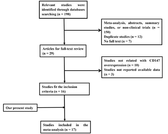
Figure 1: Flowchart showing the selection process for the included studies.
GC group vs. control group
A total of 10 studies [7, 8, 10-17] (Table 1) which reported the expression of CD147 in GC group (GC tissues) and control group (pericarcinoma tissues or normal gastric tissues) were included for this analysis. Our results indicated that expression rate of CD147 in the GC group was higher than that in the control group, and the difference between the two groups was statistically significant (OR = 12.80, 95% CI: 4.89 - 33.50, P = 0.000, I2 = 86.2%, Figure 2). Furthermore, our meta-analysis demonstrated that CD147 expressions in GC tissues were significantly higher than normal mucosa tissues (OR = 9.36, 95% CI: 2.09 - 42.00, P = 0.000, I2 = 89.3%, Figure 3A).
Table 1: Characteristics of the case–control studies included in the meta-analyses
Cases |
Controls |
||||||
Author |
Yeara |
Country |
high expression |
low expression |
high expression |
low expression |
Tissue for cases |
Zhou et al. |
2013 |
China |
37 |
33 |
0 |
5 |
Normal mucosa tissues |
Zhou et al. |
2015 |
China |
49 |
14 |
10 |
30 |
Adjacent non-tumor tissues |
Huang et al. |
2007 |
China |
40 |
18 |
0 |
20 |
Adjacent non-tumor tissues |
Pinheiro et al. |
2009 |
Portugal |
66 |
94 |
26 |
40 |
Normal mucosa tissues |
Gao et al. |
2009 |
China |
51 |
19 |
1 |
4 |
Normal mucosa tissues |
Xu et al. |
2010 |
China |
56 |
9 |
11 |
54 |
Adjacent non-tumor tissues |
He at al. |
2009 |
China |
40 |
10 |
6 |
24 |
Normal mucosa tissues |
Miao et al. |
2011 |
China |
49 |
10 |
2 |
8 |
Normal mucosa tissues |
Wu et al. |
2010 |
China |
131 |
30 |
10 |
30 |
Adjacent non-tumor tissues |
Zheng et al. |
2014 |
Japan |
551 |
445 |
6 |
104 |
Normal mucosa tissues |
a Year of publicate
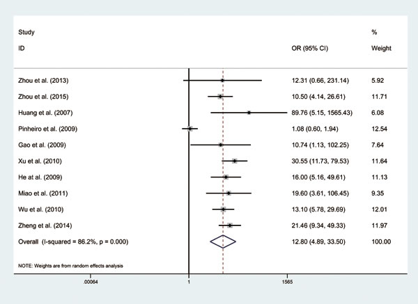
Figure 2: Forest plot of odd ratios (ORs) of 10 included studies using a random-effect model.
Subgroup analysis by gender: male group vs. female group
A total of 10 studies [8, 10-12, 17-21] (including ours) (Supplementary Table S1) involving 2305 patients were included in this subgroup. Compared with female group, expression rate of CD147 is higher in male group. A significant statistical difference was observed (OR = 1.27, 95% CI: 1.07 - 1.51, P = 0.499, I2 = 0%, Figure 3B).
TNM stage of GC tissues: high stage group vs. low stage group
A total of 12 studies [7, 8, 10-14, 16, 18, 21, 22] (including ours) (Supplementary Table S2) reported the expression of CD147 in high stage group and low stage group of GC tissues. Meta-analysis of fixed effect model showed that expression rate of CD147 in the high stage group was higher than that in low stage group. The difference between the two groups was statistically significant (OR = 3.29, 95% CI: 2.53 - 4.30, P = 0.109, I2 = 35.1%, Figure 3C).
Lymph node (LN) metastasis of GC tissues: positive group vs. negative group
A total of 14 studies [7, 8, 10-19, 21, 22] (Supplementary Table S3) reported the expression of CD147 in positive and negative lymph node metastasis of GC tissues. Our result showed that expression rate of CD147 in the positive group (LN+) is higher than that in negative group (LN−). The difference between two groups was statistically significant (OR = 3.05, 95% CI: 1.83 - 5.08, P = 0.000, I2 = 80.3%, Figure 3D).
Differentiation of GC tissues: Poor group vs. Well and Moderate group
A total of 12 studies [7, 8, 10, 11, 13, 15, 16, 18-21, 23] (Supplementary Table S4) reported the expression of CD147 in well and moderate group and poor group of GC tissues. Our results showed that expression rate of CD147 in the poor group is higher than the Well and Moderate group. There was difference between two groups (OR = 1.62, 95% CI: 1.11 - 2.35, P = 0.004, I2 = 60.0%, Figure 3E).
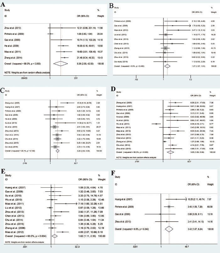
Figure 3: Forest plot of odd ratios (ORs) of subgroup analysis. A. subgroup analysis based on control tissues. B. subgroup analysis based on gender. C. subgroup analysis based on TNM stage. D. subgroup analysis based on LN metastasis. E. subgroup analysis based on differentiation. F. subgroup analysis based on depth of invasion.
Depth of invasion of GC tissues: Muscular propria/subserosa vs. Mucosa
A total of 4 studies [7, 8, 10, 12] (Supplementary Table S5) reported the expression of CD147 in Muscular propria/subserosa group and Mucosa group of GC tissues. Our result showed that expression rate of CD147 in the Muscular propria/subserosa group is higher than that in Mucosa group. The difference between two groups was statistically significant (OR = 3.42, 95% CI: 1.87 - 6.24, P = 0.349, I2 = 8.9%, Figure 3F).
Table 2: Relationship between CD147 expression and clinicopathological features of gastric cancer patients
Clinical parameter |
No. of patients |
CD147 |
P-value |
|
low(n=67) |
high(n=76) |
|||
Age |
||||
≤65 |
73 |
36 |
37 |
0.547 |
>65 |
70 |
31 |
39 |
|
Gender |
||||
male |
100 |
46 |
54 |
0.755 |
female |
43 |
21 |
22 |
|
tumor_max_diameter |
||||
d<5 |
73 |
37 |
36 |
0.348 |
d≥5 |
70 |
30 |
40 |
|
T |
||||
T1-2 |
29 |
15 |
14 |
0.556 |
T3-4 |
114 |
52 |
62 |
|
N |
||||
N0 |
37 |
24 |
13 |
0.011 |
N1-3 |
106 |
43 |
63 |
|
M |
||||
M0 |
136 |
65 |
71 |
0.448 |
M1 |
7 |
2 |
5 |
|
stage |
||||
Ⅰ+Ⅱ |
60 |
35 |
25 |
0.019 |
Ⅲ+Ⅳ |
83 |
32 |
51 |
|
Publication bias and sensitivity Analysis
The publication bias was assessed by Begg’s funnel plot and Egger’s test. The results showed there were no publication bias in the analysis (Data not shown). The stability of the study was detected by sensitivity analysis, by excluding one individual study in sequence. Our analysis displayed that result was not changed.
CD147 expression is closely associated with clinicopathological features in GC patients.
We calculated the expression levels of CD147 protein in 143 paraffin-embedded GC samples using immunohistochemical staining. The power analysis indicates our sample size is enough (Power = 0.9997). As shown in Figure 4, CD147 expression is more frequent in GC tissues than that in adjacent non-tumor tissues. The relationship between the expression level of CD147 protein and the clinicopathological parameters of GC was analyzed. As summarized in Table 2, overexpression of CD147 protein is significantly associated with clinical N stage (N0 vs. N1-N3; P = 0.011), and tumor stage (stage I-II vs. stage III-IV; P = 0.019).
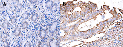
Figure 4: Expression of CD147 in GC and adjacent non-tumor tissues. A. expression of CD147 in GC tissues. B. expression of CD147 in adjacent non-tumor tissues.
Correlation between CD147 expression and clinical outcome in subtypes of GC
In order to investigate the association between CD147 expression and clinical outcome, we divided the patients into CD147-high and CD147-low groups, and survival analyses were performed using the Kaplan-Meier method. In summary, the patients with higher CD147 expression had significant lower OS (P < 0.0001) than those with lower CD147 expression (Figure 5A). Meanwhile, the results of survival analyses coming from the low stage (stage I-II) groups (P = 0.0001, Figure 5B), and the high stage (stage III- IV) groups (P = 0.001, Figure 5C) demonstrated that the OS was lower in patients with higher expression of CD147.
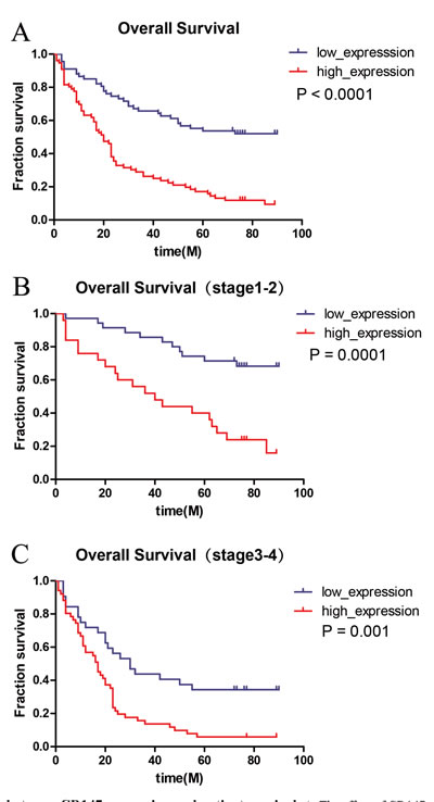
Figure 5: Association between CD147 expression and patient survival. A. The effect of CD147 expression on OS on the whole (log-rank p < 0.05). B., C. OS were significantly different between the low expression group and the high expression group both in low stage(stage I-II) and high stage(III-IV) (log-rank p < 0.05).
Univariate and Multivariate analyses of OS in patients with GC
Based on the result of univariate analysis, it demonstrated that CD147 overexpression was significantly associated with poor overall survival (OS) (HR = 3.206, 95% CI = 2.092-4.915, P = 8.95E-08). In multivariate survival analysis, T, N, and M collapsing into stage as a whole, together with age and gender, were included in the Cox proportional hazard models as adjusted variables. Significantly, overexpression of CD147 is negatively correlated with OS (HR = 2.825, 95% CI = 1.833-4.352, P = 2.51E-06), which indicated that CD147 can act as an independent predict factor for GC patients (Table 3). The other meaningful prognostic factors for OS in GC is tumor stage (HR = 2.618, 95%CI = 1.689-4.056, P = 1.66E-05) which has been universally acknowledged (Figure 6).
Table 3: Univariateand multivariate analysis of overall survival in 90 patients with gastric cancer
Factor |
Univariate analysis |
Multivariate analysis |
||
HR(95%CI) |
P |
HR(95%CI) |
P |
|
Age |
1.021(1.001-1.041) |
0.039 |
1.018(0.998-1.038) |
0.074 |
Gender |
0.928( 0.605-1.424) |
0.733 |
0.944(0.614-1.453) |
0.794 |
Stage |
2.898(1.883- 4.461) |
1.32E-06 |
2.618(1.689-4.056) |
1.66E-05 |
Expression |
3.206(2.092-4.915) |
8.95E-08 |
2.825(1.833-4.352) |
2.51E-06 |
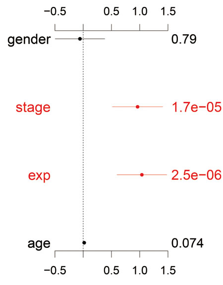
Figure 6: Multivariate analysis of overall survival. CD147 expression as an independent prognostic factor in gastric cancer.
Discussion
To the best of our knowledge, this is the first meta-analysis, which included sixteen published studies (1752 cases and 391 controls) and one original study (143 cases) from our own research, to assess the association between CD147 and the clinicopathological features of GC. Our results indicate that expression rate of CD147 in the GC group is higher than that in control group. Besides, gender, TNM stage, lymph node metastasis, and depth of invasion have a relationship with the expression state of CD147. Further, our retrospective analysis of 143 cases demonstrated CD147 protein expression can be an independent prognosis biomarker for GC with a strong statistical power.
In recent years, although surgery, chemoradiotherapy, and targeted therapy make great progress to prolong survival of GC patients, GC is still the third leading cause of cancer-related death worldwide mainly due to tumor local recurrence and distant metastasis [24]. With the development of related genomic and molecular technology, some biomarkers for GC diagnosis and treatment were discovered. However, sensitive biomarkers which are able to diagnose GC in early stage and predict prognosis of GC patients are needed.
CD147 is a 57-kilodalton (kDa) transmembrane glycoprotein which belongs to the immunoglobulin (IgG) superfamily [25]. Moreover, CD147, comprised of 2 extracellular Ig domains, is a single transmembrane domain required for counter receptor binding activity which is involved in matrix metalloproteinases (MMPs) induction and a short cytoplasmic domain that interacts with Cav 1 [26, 27]. CD147 mediates tumor cell-macrophage interactions, and has been shown to induce both MMPs and vascular endothelial growth factor (VEGF) [28]. VEGF plays an important role in angiogenesis [29]. It is notable that angiogenesis is necessary for tumor growth and metastasis and constitutes an important point in the control of cancer progression [30]. Frequently, tumors overexpress proangiogenic factors, such as vascular endothelial growth factor, allowing them to make this angiogenic switch [31].
Based on our meta-analysis, results showed that the expression rate of CD147 in the GC group was higher than that in control group. Furthermore, CD147 expressions in GC tissues were significantly higher than normal mucosa tissues. When exploring the diagnostic value of CD147 in GC by immunohistochemistry, we found that CD147 expression level was higher in GC tissues than in adjacent non-tumor tissues. Similar with our study, Xu et al. [13] and Wu et al. [16] found that CD147 expression in GC tissues was higher than that in paired adjacent non-tumor tissues. Except for GC, CD147 was found relevant in other malignancies like colorectal cancer [32], ovarian cancer [33], and clear cell renal cell carcinoma [34]. Thus, it is not surprising that the expression rate of CD147 is higher in GC tissues.
Previous studies have identified gender differences that may contribute to the development of GC [35, 36]. It may reflect that the role of biological factors is important and suggest that female might be intrinsically more vigorous than men in coping with cancer [37]. However, our research showed there was no significant statistic between male group and female group. Interestingly, the expression rate of CD147 in male group was higher than that in female group in our meta-analysis. Due to the lack of detailed source data, we cannot perform a subgroup analysis based on age. Thus, more research should be done in the future.
Invading basement membrane (BM) barriers is essential steps in the pathology of GC [38]. Depth of invasion is the most important predictors of survival [39]. The deeper the invasion into the stomach wall, the poorer the prognosis will be [40]. So, the meta-analysis showed that expression rate of CD147 in the Muscular propria/subserosa group is higher than that in Mucosa group.
The TNM Classification of Malignant Tumors (TNM) is a cancer staging notation system that gives codes to describe the stage of a person’s cancer. The higher stages represent higher progression of cancer. Previously, Wu et al. [16] indicated that CD147 was over expressed in high stage compared with low stage. Our meta-analysis also showed that expression rate of CD147 in the high stage group was higher than that in low stage group. In fact, due to the higher malignancy, it is not surprising that the high stage tissues express more CD147 than the low stage tissues. In the analysis of the association between CD147 and clinicopathological features of GC patients, our retrospective research proved that TNM stage and N stage were associated with expression of CD147. In addition, Miao et al. [19] demonstrated that overexpression of CD147 is associated with Lymph node (LN) metastasis of GC. Based on our meta-analysis, our result confirmed that expression rate of CD147 in the positive group (LN+) is higher than that in negative group (LN−). In fact, lymph node metastasis is the most common way for tumor metastasis. Even in early GC (EGC), the incidence of LN metastasis exceeds 10% [41]. GC with lymph node metastasis has more damage to patient which means more serious patient’s condition.
After analyzing the cases with clinic information in our retrospective data, we found that the OS of the GC group with low CD147 expression had a longer survival time than that of high CD147 expression group. The univariate and multivariate analyses also demonstrated that CD147 overexpression was significantly associated with poor OS. Notably, the patients with lower CD147 expression showed a longer survival time compared with high CD147 expression patients in low stage group and high stage group. This may indicate that the level of CD147 expression can be used as a molecular biomarker for the prognosis of GC. Since good prognostic prediction is necessary to ascertain the risk and the effectiveness of treatments such as surgery and chemoradiotherapy, CD147 may be a competent prognostic marker to predict treatment response.
However, several limitations about our research should be addressed. First, there are high heterogeneity in GC group vs. control group in the meta-analysis. After we excluded Pinheiro et al.’s study [12], I squared turn to 0, but the result did not changed. A possible reason is that population of this study were Portugal, which different with other studies. Second, except 3 studies published in English, the other 13 studies were all in Chinese, the language bias is unavoidable. Third, most study participants were Chinese, so the results may not be generalizable to other races. Fourth, due to the limitation of included studies, we cannot assess the role of CD147 in prediction the response of treatment. Thus, further studies with larger numbers of patients and more comprehensive clinical data are warranted to explore the full picture of the role of CD147 in GC.
Above all, CD147 upregulated in GC tissues compared with noncancerous tissues. Moreover, gender, TNM stage, lymph node metastasis, and depth of invasion are all associated with CD147. Our retrospective analysis demonstrated CD147 protein expression was significantly associated with clinical N stage, and tumor stage. Meanwhile, it can also serve as an independent prognosis biomarker. In conclusion, our results support the role of CD147 as a good indicator of diagnosis and prognosis.
materials and Methods
Meta-analysis
Literature search
Eligible publications were retrieved by searching PubMed, Embase, and Chinese National Knowledge Infrastructure (CNKI) databases up to April 22, 2016. The search strategy was based on the following words: (“antigens, cd147”[MeSH Terms] OR (“antigens”[All Fields] AND “cd147”[All Fields]) OR “cd147 antigens”[All Fields] OR “cd147”[All Fields]) AND (“stomach neoplasms”[MeSH Terms] OR (“stomach”[All Fields] AND “neoplasms”[All Fields]) OR “stomach neoplasms”[All Fields] OR (“gastric”[All Fields] AND “cancer”[All Fields]) OR “gastric cancer”[All Fields]). Furthermore, we also searched the additional publications from the reference lists of the retrieved articles or reviews which had been previously missed.
Inclusion and exclusion criteria
In this meta-analysis, publications that met the following criteria were selected as candidate articles: (1) inclusion of pathologically confirmed GC patients; (2) investigation of the relationship between the expression of CD147 and GC; (3) available data for calculation of odds ratios (ORs) with their corresponding 95% confidence intervals (CIs). And the following exclusion criteria were used: (1) abstracts and reviews, (2) insufficient data to extract or calculate the ORs, and (3) repeated or overlapping publications.
Data extraction
Two authors identified and screened the search findings. A third author reviewed all data entries. The following items were extracted from the eligible studies: name of the first author, publication year, country of the first author, sample size and numbers of cases and controls.
Statistical analysis
In this meta-analysis, all statistical analyses were performed using the STATA 12.0 software (StatCorp, College Station, TX, USA). Pooled odds ratios (ORs) and 95% confidence intervals (CIs) were calculated to evaluate the strength of the associations. We judged heterogeneity by calculating the I2 statistic, where an I2 value from 0 to 25% indicate low heterogeneity, 25-50% moderate heterogeneity and ≥ 50% high heterogeneity. If data was not significantly heterogeneous (P > 0.05 or I2 < 50 %), the pooled effects were calculated using a fixed model, Otherwise, a random-effects model was employed [42]. Publication bias was evaluated using the Egger’s and Begg’s test. We also employed sensitivity analysis to evaluate stability of the results.
Study population
Patients with GC were recruited from a study carried out between 2009 and 2014 at Taihe Hospital, Hubei University of Medicine. Clinic pathologic data for parameters were collected from the pathology report. None of the patients had received radiotherapy or chemotherapy before surgery. From their medical records the data on date of diagnosis, age, sex, tumor size, lymph node metastasis, TNM stage (TNM stages I and II were classified as low stage, III and IV as high stage), and the overall survival were extracted. The study has been approved by the institutional review board at Hubei University of Medicine. All methods used in this study were carried out in accordance with the approved guidelines and all experimental protocols were approved by Hubei University of Medicine. Informed consent was obtained from all subjects.
Immunohistochemistry
After surgical procedures, clinical tissue samples were fixed in 4% paraformaldehyde. Before cutting into 4 μm sections, the samples were embedded in paraffin. The sections were dewaxed and rinsed in 100% xylene for 10 minutes. The sections were then rehydrated in 100% alcohol, 95% alcohol, 90% alcohol, 80% alcohol and 70% alcohol for 5 minutes. Then, 5 % hydrogen peroxide was applied to block endogenous peroxide activity and the samples were washed with phosphate-buffered saline (PBS). Blocking was performed with 5% sodium citrate solution for 15 min at 100 °C. The sections were incubated at 4°C overnight with the anti-CD147 antibody (1:500) (Sigma, St. Louis, MO, USA). Next day, the slides were washed with PBS and incubated with secondary antibody for 20 minutes at 35 °C, followed by color development with Diaminobenzidine (DAB) for 3 minutes after washed with PBS. The cell nuclei were counterstained with hematoxylin for 3 minutes. Sections were photographed on an Olympus photomicroscope (Inha, Japan). The degree of IHC staining was evaluated by two independent pathologists. Staining intensity was graded as “0” (negative), “1” (weak), “2” (moderate) and “3” (strong); staining percentage was graded as “0” ( < 5%), “1” (5-25%), “2” (25-50%), “3” (50-75%) or “4” (>75%). A final immunoreactivity scores (IRS) was calculated by multiplying the values of the staining intensity and staining percentage. The IRS value > 4 was defined as high expression and IRS value ≤ 4 as low expression. ImagePro Plus (Media Cybernetics, Silver Spring, MD, USA) was used to quantitatively score the tissue sections.
Statistic method
The SPSS 19.0 (IBM Corporation, Armonk, NY, USA), GraphPad Prism Software 5 (GraphPad, Inc.; La Jolla; California; USA) and R software (R3.3.0) for windows was used for statistical analysis. Correlation between CD147 expression and clinicopathological characteristics were evaluated by Chi-square test and Fisher’s exact tests. Overall survival (OS) rate were estimated by using the Kaplan-Meier method and the log-rank test was used to calculate P Values. Univariate and multivariate Cox regression models were used to calculate hazard ratios and their confidence intervals for the study variables. A one-sided power calculation was performed on the effect size (hazard ratio) of CD147 expression in Cox regression [43]. Historically, the value of 0.80 (Beta = 0.20) was used for power. All the other analyses were considered statistically significant when a two-sided P < 0.05.
Abbreviations
Gastric cancer (GC); cluster of differentiation 147 (CD147); kilodalton (kDa); immunoglobulin (IgG); basement membrane (BM); phosphate-buffered saline (PBS); Diaminobenzidine (DAB); odds ratios (ORs); confidence intervals (CIs); early GC (EGC).
Acknowledgments
The authors gratefully acknowledge the Natural Science Foundation of of Hubei Provincial Department of Education (grant number: Q20162115); Shiyan Taihe Hospital youth team project (grants number: 2006TD008, 2012TD01); The Key Discipline Project of Hubei Province (grant number: 2012ZDFX04); the Initial Project for Post-Graduates of Hubei University of Medicine (grant number: 2016QDJZR10).
Conflict of Interests
The authors declare no conflict of interest.
References
1. Jemal A, Bray F, Center MM, Ferlay J, Ward E, Forman D. Global cancer statistics. CA Cancer J Clin. 2011; 61:69–90.
2. Siegel RL, Miller KD, Jemal A. Cancer statistics, 2015. CA Cancer J Clin. 2015; 65:5–29.
3. Rocco A, Nardone G. Diet, H pylori infection and gastric cancer: evidence and controversies. World J Gastroenterol. 2007; 13:2901–12.
4. He S, Liao G, Liu Y, Huang L, Kang M, Chen L. Overexpression of STAT3/pSTAT3 was associated with poor prognosis in gastric cancer: a meta-analysis. Int J Clin Exp Med. 2015; 8:20014–23.
5. Li X, Yu X, Dai D, Song X, Xu W. The altered glucose metabolism in tumor and a tumor acidic microenvironment associated with extracellular matrix metalloproteinase inducer and monocarboxylate transporters. Oncotarget. 2016; 7:23141-55. doi: 10.18632/oncotarget.8153.
6. Riethdorf S, Reimers N, Assmann V, Kornfeld JW, Terracciano L, Sauter G, Pantel K. High incidence of EMMPRIN expression in human tumors. Int J Cancer. 2006; 119:1800–10.
7. Huang Y, Zhao J, Fan K. The expression and significance of MMP-2 and CD147 in gastric cancer. Journal of Basic and Clinical Oncology. 2007; 20:21–23.
8. Gao J, Chen H, Xue J, Zhu R. Correlation of the expression of EMMPRIN and HER-2 proteins with the invasiveness and metastasis of human gastric carcinoma. World Chin J Digestology. 2009; 17:1952–56.
9. Haidich AB. Meta-analysis in medical research. Hippokratia. 2010; 14:29–37.
10. Zhou MH. The relationship between the expression of CD147 and HER-2 and invasion of gastric cancer. J Clin Exp Pathol. 2013; 29:2.
11. Zhou P, Fu Y, Gao W, Lu N. CD147 expression and HER2 in gastric cancer and its Prognostic significance. The Journal of Practical Medicine. 2015; 31:3018–21.
12. Pinheiro C, Longatto-Filho A, Simões K, Jacob CE, Bresciani CJ, Zilberstein B, Cecconello I, Alves VA, Schmitt F, Baltazar F. The prognostic value of CD147/EMMPRIN is associated with monocarboxylate transporter 1 co-expression in gastric cancer. Eur J Cancer. 2009;45:2418-24.
13. Xu J. D T, Tian X, Chen J, Wang M. Expression of PTEN and CD147 and AKT2 in gastric carcinoma tissues and the clinical significance. Experimental Research. 2010; 13:681–84.
14. He C, Zhong Y, Chen J. Expression and significace of CD147 and MMPs in gastric carcinoma. Shandong Yiyao. 2009; 49:12–13.
15. Miao J. Expression and significance of CD147 and MT1-MMP in gastric cancer. Shandong Yiyao. 2011; 51:52–53.
16. W Qian, Z Hong, Z Heping. Investigation of CD147 and MMP-2 expressionin gastric carcinoma and their significance by tissue microarra. Acta Universitatis Medicinalis Anhu. 2010; 45:43–46.
17. Zheng X, Zheng H, Zhao E, Xiao L, Zhao S, et al. Expression and significance of CD147 protein in gastrointestinal cancer. Journal of chengde medical college. 2014;31:187-9.
18. Chen T, Xie L, Zhou X. CD147 And the integrin α3 Expression and Significance in Gastric carcinoma. Shandong Yiyao. 2013; 53:33–34.
19. Miao J. Expression of HER-2, mt1-mmp and cd147 in gastric carcinoma and their significances. J Clin Exp Pathol. 2012; 28:1361–64.
20. Liu L, Guo L, Wang Z, Ji J, Zhang J, et al. Expression of HAb18G/CD147 in large sample of gastric cancer tissue specimen. J Diagn Concepts Pract. 2012; 11:42–46.
21. Chu D, Zhu S, Li J, Ji G, Wang W, Wu G, Zheng J. CD147 expression in human gastric cancer is associated with tumor recurrence and prognosis. PLoS One. 2014; 9:e101027.
22. Zheng X, Ma H, Wu Q, Huang Y, Wu J. Expression of CD147 in gastric carcinoma and its clinical significance. CHIN J CANCER PREV TREAT. 2005; 12:1385–87.
23. Zheng HC, Takahashi H, Murai Y, Cui ZG, Nomoto K, Miwa S, Tsuneyama K, Takano Y. Upregulated EMMPRIN/CD147 might contribute to growth and angiogenesis of gastric carcinoma: a good marker for local invasion and prognosis. Br J Cancer. 2006; 95:1371–78.
24. Zhang CX, Wang SY, Chen SQ, Yang SL, Wan L, Xiong B. Association between pretreatment Glasgow prognostic score and gastric cancer survival and clinicopathological features: a meta-analysis. Onco Targets Ther. 2016; 9:3883–91.
25. Gabison EE, Mourah S, Steinfels E, Yan L, Hoang-Xuan T, Watsky MA, De Wever B, Calvo F, Mauviel A, Menashi S. Differential expression of extracellular matrix metalloproteinase inducer (CD147) in normal and ulcerated corneas: role in epithelio-stromal interactions and matrix metalloproteinase induction. Am J Pathol. 2005; 166:209–19.
26. Yu XL, Hu T, Du JM, Ding JP, Yang XM, Zhang J, Yang B, Shen X, Zhang Z, Zhong WD, Wen N, Jiang H, Zhu P, Chen ZN. Crystal structure of HAb18G/CD147: implications for immunoglobulin superfamily homophilic adhesion. J Biol Chem. 2008; 283:18056–65.
27. Luo J, Teplyakov A, Obmolova G, Malia T, Wu SJ, Beil E, Baker A, Swencki-Underwood B, Zhao Y, Sprenkle J, Dixon K, Sweet R, Gilliland GL. Structure of the EMMPRIN N-terminal domain 1: dimerization via beta-strand swapping. Proteins. 2009; 77:1009–14.
28. Walter M, Simanovich E, Brod V, Lahat N, Bitterman H, Rahat MA. An epitope-specific novel anti-EMMPRIN polyclonal antibody inhibits tumor progression. OncoImmunology. 2016; 5:e1078056.
29. Ohta Y, Shridhar V, Bright RK, Kalemkerian GP, Du W, Carbone M, Watanabe Y, Pass HI. VEGF and VEGF type C play an important role in angiogenesis and lymphangiogenesis in human malignant mesothelioma tumours. Br J Cancer. 1999; 81:54–61.
30. Folkman J. The role of angiogenesis in tumor growth. Semin Cancer Biol. 1992; 3:65–71.
31. Folkman J. Role of angiogenesis in tumor growth and metastasis. Semin Oncol. 2002; 29:15–18.
32. Zhu S, Chu D, Zhang Y, Wang X, Gong L, Han X, Yao L, Lan M, Li Y, Zhang W. EMMPRIN/CD147 expression is associated with disease-free survival of patients with colorectal cancer. Med Oncol. 2013; 30:369.
33. Gao J, Hu Z, Liu J, Liu D, Wang Y, Cai M, Zhang D, Tan M, Lin B. Expression of CD147 and Lewis y antigen in ovarian cancer and their relationship to drug resistance. Med Oncol. 2014; 31:920.
34. Kim Y, Choi JW, Lee JH, Kim YS. Expression of lactate/H () symporters MCT1 and MCT4 and their chaperone CD147 predicts tumor progression in clear cell renal cell carcinoma: immunohistochemical and The Cancer Genome Atlas data analyses. Hum Pathol. 2015; 46:104–12.
35. Forman D, Burley VJ. Gastric cancer: global pattern of the disease and an overview of environmental risk factors. Best Pract Res Clin Gastroenterol. 2006; 20:633–49.
36. Compare D, Rocco A, Nardone G. Risk factors in gastric cancer. Eur Rev Med Pharmacol Sci. 2010; 14:302–08.
37. Somi MH, Ghojazadeh M, Bagheri M, Tahamtani T. Clinicopathological factors and gastric cancer prognosis in the iranian population: a meta-analysis. Asian Pacific journal of cancer prevention. 2015; 16:853–57.
38. Lukaszewicz-Zajac M, Mroczko B, Szmitkowski M. Gastric cancer - The role of matrix metalloproteinases in tumor progression. Clinica chimica acta. 2011;412:1725-30.
39. Martin RC 2nd, Jaques DP, Brennan MF, Karpeh M. Extended local resection for advanced gastric cancer: increased survival versus increased morbidity. Ann Surg. 2002; 236:159–65.
40. Tsujitani S, Kakeji Y, Watanabe A, Kohnoe S, Maehara Y, Sugimachi K. Infiltration of dendritic cells in relation to tumor invasion and lymph node metastasis in human gastric cancer. Cancer. 1990; 66:2012–16.
41. Roviello F, Rossi S, Marrelli D, Pedrazzani C, Corso G, Vindigni C, Morgagni P, Saragoni L, de Manzoni G, Tomezzoli A. Number of lymph node metastases and its prognostic significance in early gastric cancer: a multicenter Italian study. J Surg Oncol. 2006; 94:275–80.
42. Xiao F, Lan A, Lin Z, Song J, Zhang Y, Li J, Gu K, Lv B, Zhao D, Zeng S, Zhang R, Zhao W, Pan Z, et al. Impact of CAG repeat length in the androgen receptor gene on male infertility - a meta-analysis. Reprod Biomed Online. 2016; 33:39–49.
43. Hsieh FY, Lavori PW. Sample-size calculations for the Cox proportional hazards regression model with nonbinary covariates. Control Clin Trials. 2000; 21:552–60.