
INTRODUCTION
In 2012, nearly 8.2 million people reportedly died from cancer and about 14.1 million people were diagnosed with cancer worldwide [1]. The American National Center for Health Statistics has estimated that nearly 600 thousand Americans will die of cancer in 2016 [2]. The five year survival rate of most cancers is extremely low and since survival depends on early diagnosis of cancer, there is a constant need to identify and develop newer diagnostic and prognostic markers.
Long noncoding RNA (lncRNA) are transcribed RNA molecules that lack an open reading frame and are longer than 200 nucleotides [3]. They are involved in epigenetic regulation, transcriptional and posttranscriptional regulation that are key cellular processes that also determine tumorigenesis [4]. Dysregulation of lncRNAs has been reported in many types of cancers [5–8]. Since they have been implicated in different stages of cancer progression including proliferation, invasion and metastasis, they are promising prognostic markers for cancer [9–10, 11]. Due to the specific expression of lncRNA in the development of tumor and their presence in body fluids and tumor tissues, they are promising biomarkers to diagnose and monitor tumors [12]. Therefore, identification of tumor related lncRNAs that are vital in tumorigenesis are promising biomarkers for cancer prognosis.
HULC (Highly Upregulated in Liver Cancer) was first reported in liver cancer and showed extensive regulatory functions in cell proliferation, apoptosis, invasion, cell cycle, and drug resistance [13]. HULC is an lncRNA with two non-translated exons and about 500 nucleotides long. It is highly expressed in hepatocellular carcinoma and colorectal cancer with liver metastasis [14]. Since HULC expression has been shown during cancer growth and metastasis, it is a promising prognostic biomarker candidate for human cancers [15]. In recent years, HULC has been found to be dysregulated in osteosarcoma, pancreatic cancer, colorectal cancer, hepatocellular carcinoma, gastric cancer, hepatocellular carcinoma and large B-cell lymphoma [16–22]. Du and others found that the HULC promoted liver cancer cell proliferation by inhibiting P18 [23]. Silencing of HULC effectively reversed EMT phenotype in gastric cancer [24]. These studies revealed that HULC has potential prognostic value in cancer patients. However, most studies regarding HULC are limited by discrete outcomes and small patient samples. Therefore, we performed this meta-analysis to determine the prognostic value of HULC by combined analysis of data from multiple studies.
RESULTS
Literature search analysis results
The detailed screening process of HULC studies is shown in Figure 1. Based on the inclusion and exclusion criteria, a total of seven studies and 730 patients were included in the meta-analysis [16–22]. The characteristics of the seven studies are summarized in Table 1. The total number of subjects analyzed in the seven studies ranged from 33 to 304, with a mean sample size of 104.3. Six of the seven studies were conducted in China whereas one study was from Brazil and were published between 2014 and 2016. Among the seven studies, two focused on osteosarcoma (OSC) [20–21] and one each on gastric cancer [16], hepatocellular carcinoma [17], pancreatic cancer [18], large B-cell lymphoma [19] and colorectal cancer [22]. HULC expression was measured in the tumor specimen as well as the serum. The diagnosis of LNM, DM and tumor stage depended on the pathology. The NOS scores of all the studies were ≥7.
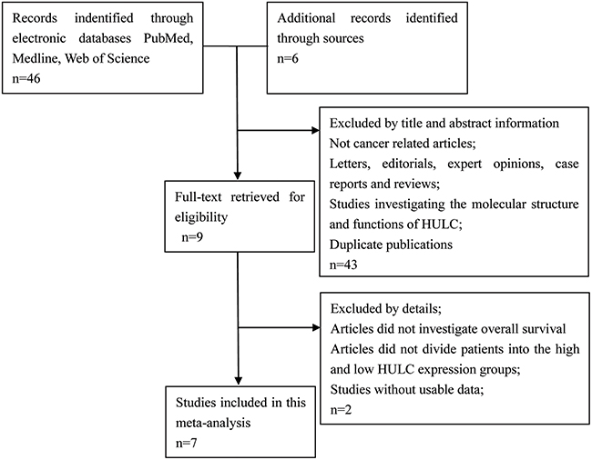
Figure 1: Flowchart showing the steps of literature search and selection criteria for the meta-analysis.
Table 1: The basic information and data of all included studies in the meta-analysis
Study |
Year |
Region |
Tumor type |
Age |
Men % |
Reference gene |
Sample size |
HULC expression |
Analysis (OS) |
HR(95% CI) |
NOS |
Method |
|||||
|---|---|---|---|---|---|---|---|---|---|---|---|---|---|---|---|---|---|
Low |
High |
||||||||||||||||
Total |
LNM |
DM |
Total |
LNM |
DM |
||||||||||||
Jin[16] |
2016 |
China |
GC |
60 |
65 |
GAPDH |
100 |
52 |
25 |
2 |
48 |
34 |
9 |
Multivariate |
0.58(0.23-1.44) |
8 |
qRT-PCR |
Li[17] |
2016 |
China |
HCC |
- |
76.3 |
GAPDH |
38 |
15 |
- |
1 |
23 |
- |
8 |
Multivariate |
0.47(0.11-2.01) |
8 |
qRT-PCR |
Peng[18] |
2014 |
China |
PC |
- |
55.3 |
GAPDH |
304 |
92 |
23 |
- |
212 |
157 |
- |
Multivariate |
0.352(0.172-0.752) |
7 |
qRT-PCR |
Peng[19] |
2016 |
China |
LBCL |
- |
70.4 |
GAPDH |
142 |
47 |
- |
- |
95 |
- |
- |
Multivariate |
0.738(0.414-1.288) |
7 |
qRT-PCR |
Sun[20] |
2015 |
China |
OSC |
- |
57.7 |
GAPDH |
78 |
39 |
- |
5 |
39 |
- |
16 |
Multivariate |
0.439(0.184-0.675) |
8 |
qRT-PCR |
Uzan[21] |
2016 |
Brazil |
OSC |
5 |
48.5 |
GAPDH |
33 |
21 |
- |
- |
12 |
- |
- |
Multivariate |
0.045(0.0046–0.443) |
7 |
qRT-PCR |
Yang[22] |
2016 |
China |
CRC |
- |
- |
GAPDH |
35 |
12 |
- |
- |
23 |
- |
- |
Multivariate |
0.43 (0.05-4.04) |
8 |
qRT-PCR |
Note: The dashes represent no data.
Abbreviations: GAPDH, glyceraldehyde-3-phosphate dehydrogenase; GC, gastric cancer; HCC, hepatocellular carcinoma; PC, pancreatic cancer; LBCL, large B-cell lymphoma; OSC, osteosarcoma; CRC, colorectal cancer; LNM, lymph node metastasis; DM, distant metastasis.
Association between HULC expression levels and OS
We performed cumulative meta-analysis to determine the role of HULC in overall survival (OS) of all 730 cancer patients from the seven studies. Statistical analyses revealed that HULC was associated with OS of cancer patients (pooled HR= 0.50, 95% CI: 0.35–0.70, P <0.00001; Figure 2). Our analyses did not find any significant heterogeneity among the studies (I2=16%, PQ=0.31). Therefore, our data demonstrated that HULC was an independent OS factor among cancer patients and its high expression was associated with shorter OS.
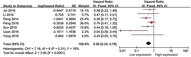
Figure 2: Forest plot showing association between OS and elevated HULC expression in the different types of cancer.
Association between HULC expression level and LNM
Data of 404 cancer patients from 2 eligible studies was collected and analyzed. The random effects model was used due to significant heterogeneity (I2=81%, P=0.02) and the odds ratio (OR) determined as 0.20 (95% CI: 0.06–0.64, P=0.007; Figure 3). Although our analysis demonstrated significant differences in the LNM incidence between the two groups, patients with higher expression of HULC were more prone to developing LNM.
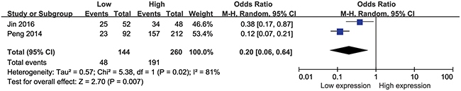
Figure 3: Forest plot showing association between HULC expression levels and lymph node metastasis.
Association between HULC expression levels and DM
Analysis of 216 patients from three eligible studies demonstrated association between HULC expression levels and the number of cancer patients with DM (Figure 4). Analysis by the fixed effects model showed no significant heterogeneity (I2=0%, PQ=0.84) and the pooled OR was 0.27 (95% CI: 0.13–0.54, P=0.0003; Figure 4). These results indicated that patients with high HULC expression level in the tumor tissues may have increased probability of DM.
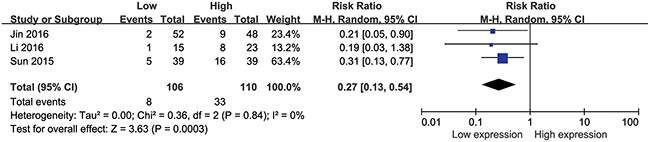
Figure 4: Forest plot showing association between HULC expression levels and distant metastasis.
Association between HULC expression levels and tumor stage
A total of 391 patients from five eligible studies were analyzed for the relationship between the HULC expression levels and the tumor stage in this meta-analysis. Our data demonstrated that higher the HULC expression, higher was the tumor grade with a pooled OR of 0.39 (95% CI: 0.25–0.59, P <0.0001; Figure 5) with no obvious heterogeneity (I2=16%, PQ=0.31). Therefore, our results demonstrated that higher expression of HULC significantly increased the risk of high tumor stage.
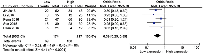
Figure 5: Forest plot showing meta-analysis of the role of HULC on tumor stage in the different types of cancer.
Begg’s funnel plot analysis
We did not find any obvious asymmetry for either overall survival (Figure 6) or tumor stage (Figure 7) when we used Begg’s funnel plot to analyze publication bias. Therefore, our findings were due to a relationship between HULC expression and the pathological parameters analyzed and not due to publication bias.
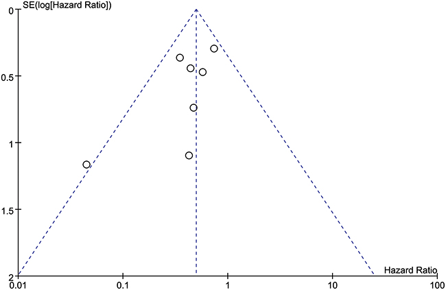
Figure 6: Funnel plot analysis to determine publication bias for the independent role of HULC on OS in the different types of cancers.
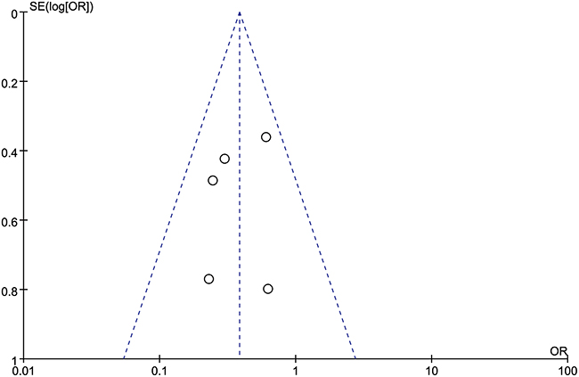
Figure 7: Funnel plot analysis to determine publication bias for the independent role of HULC on tumor stage in the different types of cancers.
DISCUSSION
Cancer is a major threat to human health all over the world and the incidence of cancer has increased gradually over the years [2]. Most cancers eventually metastasize as lymph node metastasis (LNM) and distant metastasis (DM). The occurrence of metastasis indicates poor prognosis and hence is an important indicator for survival. [25–26]. Moreover, LNM and DM are important for the diagnosis of TNM (tumor–node–metastasis) staging for cancer patients, as well as important indicators for predicting prognosis. Since the precise mechanism on metastasis remains unknown in most cancers, molecular biomarkers play a critical role in the diagnosis, prognosis and treatment of cancer [27–28]. Therefore finding new molecular markers that accurately predict tumor metastasis are of paramount importance.
Since the role of HULC as a molecular biomarker in human cancer was unclear, our study explored the prognostic value of HULC in cancer patients using the meta-analysis.
A random effects model or fixed effects model was used to analyze the data based on the results of heterogeneity analysis. Our data showed that higher HULC expression was indicative of advanced cancer and highlighted poor prognosis. By combining HRs from Cox multivariate analyses, there was a significant difference in OS between high and low HULC expression level groups (pooled HR 0.50, 95%CI: 0.35–0.70). In addition, we showed that higher HULC expression was associated with lymph node metastasis, distant metastasis and tumor stage. We found that high HULC expression was significantly associated with advanced tumor stages without obvious heterogeneity in different types of cancer. Due to lack of literature we could not perform meta-analysis to find if higher HULC expression in tumor issues may be related to the recurrence time [17], progression–free survival (PFS) [19] and event-free survival (EFS) [21]. We also found that HULC is a promising marker for diagnosis in tumor [24]. In gastric cancer, a receiver operating characteristic (ROC) curve was constructed to assess the diagnostic utility of serum HULC and the area under the ROC curve was determined to be 0.888, which was higher than the well known tumor markers for gastrointestinal malignancies, CA72-4 and CEA [16]. In addition, Peng and others found that the area under the ROC curve was 0.977 in pancreatic cancer [18] and 0.9765 in large B-cell lymphoma [19]. Therefore, HULC can be considered an independent diagnostic marker in cancer patients.
Nevertheless, there are few limitations that must be taken into account while interpreting the conclusions of our meta-analysis. First, most studies were from China, and only one study was from Brazil. Therefore our data may not represent globally. Second, the included type and number of cancers were small. Third, the criterion for high expression varied for different studies. Therefore, further well-designed and high-quality studies are needed to confirm the function of HULC in various cancers.
In conclusion, since high levels of HULC expression in multiple cancers are associated with poor OS, LNM, DM and tumor stage, it is a promising prognostic biomarker in cancer.
MATERIALS AND METHODS
Literature search to identify relevant studies for meta-analysis
A systematic search of multiple electronic databases, Medline, Pubmed, OVID, and Web of Science, was performed independently by two authors, Yanghua Fan and Lei Wu in accordance with the standard guidelines of meta-analysis. They searched literature from inception until August 14, 2016 for articles that reported HULC as a probable prognostic marker for survival of cancer patients [29–30]. The searches were performed by both the text word and MeSH strategy and included terms like ‘HULC’, ‘Highly upregulated in liver cancer’, ‘hepatocellular carcinoma up-regulated long non-coding RNA’, ‘lncRNA’, ‘noncoding RNA’,‘long intergenic noncoding RNA’, ‘carcinoma’, ‘neoplasm’, ‘tumor’, ‘cancer’, ‘prognostic’, ‘prognosis’, ‘outcome’, ‘survival’ or ‘recurrence’. The strategy was adjusted in different databases to maximize finding the appropriate articles. Manual searches were also performed using the reference lists of the relevant articles to retrieve eligible studies for inclusion in meta-analysis.
Selection criteria for including studies in meta-analysis
The two researchers, Yanghua Fan and Minhua Ye, independently evaluated all the data in the articles to select relevant studies for meta-analysis. The criteria used to include studies in the meta-analysis were as follows: 1) The relationship between HULC expression and survival was measured in multiple human tumors; 2) The expression level of HULC was measured either in human tumor tissue or serum and the patients were grouped according to the expression levels of HULC; 3) All tumors were confirmed by pathological or histological examinations and the pathological parameters like LNM, DNM, tumor stage were described; 4) Studies described statistical information on overall survival (OS) such as hazard ratio (HR) and 95% confidence interval (CI).
The criteria to exclude studies from the meta-analysis included: 1) The articles that were reviews, letters, editorials, case reports and expert opinions; 2) Non-English language and non-human studies; 3) Studies lacking data listed in the criteria for included studies; 4) Basic characterization studies of HULC.
Data extraction from relevant studies for meta-analysis
The two reviewers, Yanghua Fan and Lei Wu, independently extracted and examined the data from the selected original articles. Any disagreements in the literature assessment were resolved through consensus with a third reviewer, Xingen Zhu. The following details were collected from each of the study: surname of the first author, publication year, country, tumor type, median age of patients, percentage of male, reference gene, sample size, the number of patients with lymph node metastasis and distant metastasis, HR and 95% CI of elevated HULC for OS, the Newcastle-Ottawa Scale (NOS) score and the detection method of HULC.
The study quality was assessed in accordance with the Newcastle-Ottawa Scale (NOS). A total of nine items, each of which was assigned a score of 1, were measured in each study. The total scores for different studies ranged from 0 to 9. If the score was ≥7, the study was considered to be of high quality.
Statistical analysis
The statistical analysis was performed by RevMan version 5.3 software. The heterogeneity among different studies was measured by the Q and I2 tests. A probability value of I2 ≥50% and P < 0.1 indicated the existence of significant heterogeneity [31]. A random effects model or fixed effects model was selected based on the results of heterogeneity analysis. The random-effects model was used if there was significant heterogeneity among the studies or else, the fixed effects model was used. The potential publication bias was assessed by the Begg’s funnel plot. Pooled HRs and ORs were obtained from the published data. We used the HRs and 95% CIs reported in a publication when it was available and when they were not reported, the HR values were estimated from the survival information obtained from Kaplan-Meier curve. Overall survival was calculated using the log HR and standard error (SE) values [32]. Odds ratios (ORs) and their 95 % CIs were used to assess the association between HULC expression and the tumor parameters, including LNM, DM and tumor stage.
ACKNOWLEDGMENTS
This article was supported by National Natural Science Foundation of China (No.81660420 and No. 81560411), the Youth Science Foundation project of Jiangxi Province (NO. 20161BAB215253).
CONFLICTS OF INTEREST
The authors declare that there is no conflicts of interest regarding the publication of this paper.
REFERENCES
1. Ferlay J, Soerjomataram I, Dikshit R, Eser S, Mathers C, Rebelo M, Parkin DM, Forman D, Bray F. Cancer incidence and mortality worldwide: sources, methods and major patterns in GLOBOCAN 2012. Int J Cancer. 2015; 136: E359-E386.
2. Siegel RL, Miller KD, Jemal A. Cancer statistics, 2016. CA Cancer J Clin. 2016; 66: 7-30.
3. Johnsson P, Lipovich L, Grander D, Morris KV. Evolutionary conservation of long non-coding RNAs; sequence, structure, function. Biochim Biophys Acta. 2014; 1840: 1063-1071.
4. Guo H, Wu L, Yang Q, Ye M, Zhu X. Functional linc-POU3F3 is overexpressed and contributes to tumorigenesis in glioma. Gene. 2015; 554: 114-119.
5. Gu W, Gao T, Sun Y, Zheng X, Wang J, Ma J, Hu X, Li J, Hu M. LncRNA expression profile reveals the potential role of lncRNAs in gastric carcinogenesis. Cancer Biomark. 2015; 15: 249-258.
6. Zhou Y, Wang DL, Pang Q. Long noncoding RNA SPRY4-IT1 is a prognostic factor for poor overall survival and has an oncogenic role in glioma. Eur Rev Med Pharmacol Sci. 2016; 20: 3035-3039.
7. Cheng N, Li X, Zhao C, Ren S, Chen X, Cai W, Zhao M, Zhang Y, Li J, Wang Q, Zhou C. Microarray expression profile of long non-coding RNAs in EGFR-TKIs resistance of human non-small cell lung cancer. Oncol Rep. 2015; 33: 833-839.
8. Zhao QS, Li L, Zhang L, Meng XW, Li LL, Ge XF, Li ZP. Over-expression of lncRNA SBF2-AS1 is associated with advanced tumor progression and poor prognosis in patients with non-small cell lung cancer. Eur Rev Med Pharmacol Sci. 2016; 20: 3031-3034.
9. Loewen G, Jayawickramarajah J, Zhuo Y, Shan B. Functions of lncRNA HOTAIR in lung cancer. J Hematol Oncol. 2014; 7: 90.
10. Iguchi T, Uchi R, Nambara S, Saito T, Komatsu H, Hirata H, Ueda M, Sakimura S, Takano Y, Kurashige J, Shinden Y, Eguchi H, Sugimachi K, Maehara Y, Mimori K. A long noncoding RNA, lncRNA-ATB, is involved in the progression and prognosis of colorectal cancer. Anticancer Res. 2015; 35: 1385-1388.
11. Du Z, Fei T, Verhaak RG, Su Z, Zhang Y, Brown M, Chen Y, Liu XS. Integrative genomic analyses reveal clinically relevant long noncoding RNAs in human cancer. Nat Struct Mol Biol. 2013; 20: 908-913.
12. Qi P, Du X. The long non-coding RNAs, a new cancer diagnostic and therapeutic gold mine. Mod Pathol. 2013; 26: 155-165.
13. Matouk IJ, Abbasi I, Hochberg A, Galun E, Dweik H, Akkawi M. Highly upregulated in liver cancer noncoding RNA is overexpressed in hepatic colorectal metastasis. Eur J Gastroenterol Hepatol. 2009; 21: 688-692.
14. Panzitt K, Tschernatsch MM, Guelly C, Moustafa T, Stradner M, Strohmaier HM, Buck CR, Denk H, Schroeder R, Trauner M, Zatloukal K. Characterization of HULC, a novel gene with striking up-regulation in hepatocellular carcinoma, as noncoding RNA. Gastroenterology. 2007; 132: 330-342.
15. Ma Y, Huang D, Yang F, Tian M, Wang Y, Shen D, Wang Q, Chen Q, Zhang L. Long Noncoding RNA Highly Upregulated in Liver Cancer Regulates the Tumor Necrosis Factor-alpha-Induced Apoptosis in Human Vascular Endothelial Cells. DNA Cell Biol. 2016; 35: 296-300.
16. Jin C, Shi W, Wang F, Shen X, Qi J, Cong H, Yuan J, Shi L, Zhu B, Luo X, Zhang Y, Ju S. Long non-coding RNA HULC as a novel serum biomarker for diagnosis and prognosis prediction of gastric cancer. Oncotarget. 2016; 7:51763-51772. doi: 10.18632/oncotarget.10107.
17. Li SP, Xu HX, Yu Y, He JD, Wang Z, Xu YJ, Wang CY, Zhang HM, Zhang RX, Zhang JJ, Yao Z, Shen ZY. LncRNA HULC enhances epithelial-mesenchymal transition to promote tumorigenesis and metastasis of hepatocellular carcinoma via the miR-200a-3p/ZEB1 signaling pathway. Oncotarget. 2017; 8:8226-8238. doi: 10.18632/oncotarget.14153.
18. Peng W, Gao W, Feng J. Long noncoding RNA HULC is a novel biomarker of poor prognosis in patients with pancreatic cancer. Med Oncol. 2014; 31: 346.
19. Peng W, Wu J, Feng J. Long noncoding RNA HULC predicts poor clinical outcome and represents pro-oncogenic activity in diffuse large B-cell lymphoma. Biomed Pharmacother. 2016; 79: 188-193.
20. Sun XH, Yang LB, Geng XL, Wang R, Zhang ZC. Increased expression of lncRNA HULC indicates a poor prognosis and promotes cell metastasis in osteosarcoma. Int J Clin Exp Pathol. 2015; 8: 2994-3000.
21. Uzan VR, Lengert A, Boldrini E, Penna V, Scapulatempo-Neto C, Scrideli CA, Filho AP, Cavalcante CE, de Oliveira CZ, Lopes LF, Vidal DO. High Expression of HULC Is Associated with Poor Prognosis in Osteosarcoma Patients. PLoS One. 2016; 11: e156774.
22. Yang X J, Huang C Q, Peng C W, et al. Long noncoding RNA HULC promotes colorectal carcinoma progression through epigenetically repressing NKD2 expression[J]. Gene. 2016; 30: 172-178.
23. Du Y, Kong G, You X, Zhang S, Zhang T, Gao Y, Ye L, Zhang X. Elevation of highly up-regulated in liver cancer (HULC) by hepatitis B virus X protein promotes hepatoma cell proliferation via down-regulating p18. J Biol Chem. 2012; 287: 26302-26311.
24. Zhao Y, Guo Q, Chen J, Hu J, Wang S, Sun Y. Role of long non-coding RNA HULC in cell proliferation, apoptosis and tumor metastasis of gastric cancer: a clinical and in vitro investigation. Oncol Rep. 2014; 31: 358-364.
25. Cho JH, Lee YS, Sun DI, Kim MS, Cho KJ, Nam IC, Kim CS, Kim SY, Park YH, Joo YH. Prognostic impact of lymph node micrometastasis in oral and oropharyngeal squamous cell carcinomas. Head Neck. 2016; 38:E1777-E1782.
26. Li P, Wu F, Zhao H, Dou L, Wang Y, Guo C, Wang G, Zhao D. Analysis of the factors affecting lymph node metastasis and the prognosis of rectal neuroendocrine tumors. Int J Clin Exp Pathol. 2015; 8: 13331-13338.
27. Mirzaei H, Gholamin S, Shahidsales S, Sahebkar A, Jaafari MR, Mirzaei HR, Hassanian SM, Avan A. MicroRNAs as potential diagnostic and prognostic biomarkers in melanoma. Eur J Cancer. 2016; 53: 25-32.
28. Fan YH, Ye MH, Wu L, Lv SG, Wu MJ, Xiao B, Liao CC, Ji QK, Chai Y, Zhu XG. Overexpression of miR-98 inhibits cell invasion in glioma cell lines via downregulation of IKKepsilon. Eur Rev Med Pharmacol Sci. 2015; 19: 3593-3604.
29. Fan YH, Fang H, Ji CX, Xie H, Xiao B, Zhu XG. Long noncoding RNA CCAT2 can predict metastasis and poor prognosis: A meta-analysis. Clin Chim Acta. 2017. doi: 10.1016/j.cca.2017.01.016.
30. Altman DG, McShane LM, Sauerbrei W, Taube SE. Reporting Recommendations for Tumor Marker Prognostic Studies (REMARK): explanation and elaboration. PLoS Med. 2012; 9: e1001216.
31. Higgins JP, Thompson SG. Quantifying heterogeneity in a meta-analysis. Stat Med. 2002; 21: 1539-1558.
32. Fan YH, Ye MH, Wu L, Wu MJ, Lu SG, Zhu XG. BRAF-activated lncRNA predicts gastrointestinal cancer patient prognosis: a meta-analysis. Oncotarget. 2017; 8:6295-6303. doi: 10.18632/oncotarget.14061.