
Corrections:
Correction: β2-spectrin depletion impairs DNA damage repair
Metrics: PDF 224 views
1Department of Radiation Oncology, Houston Methodist Research Institute, Houston, TX, USA 2Department of Radiation Oncology, University of Texas Southwestern Medical School, Dallas, TX, USA 3Department of Microbiology and Molecular Genetics, McGovern Medical School, University of Texas Health Science Center at Houston, Houston, TX, USA
Published: April 24, 2026
This article has been corrected:It was identified that a portion of Figure 6B was improperly placed, resulting in an overlap between two images (Sptbn1−/− and Sptbn1+/+) for new DNA replication forks in MEF cells without Hydroxyurea treatment. The authors provided a revised Figure 6B, replacing the image for Sptbn1−/− cells without Hydroxyurea treatment with the correct data from the original experiments. This correction does not alter the article’s conclusions.
Original article: Oncotarget. 2016; 7:33557-33570. DOI: https://doi.org/10.18632/oncotarget.9677.
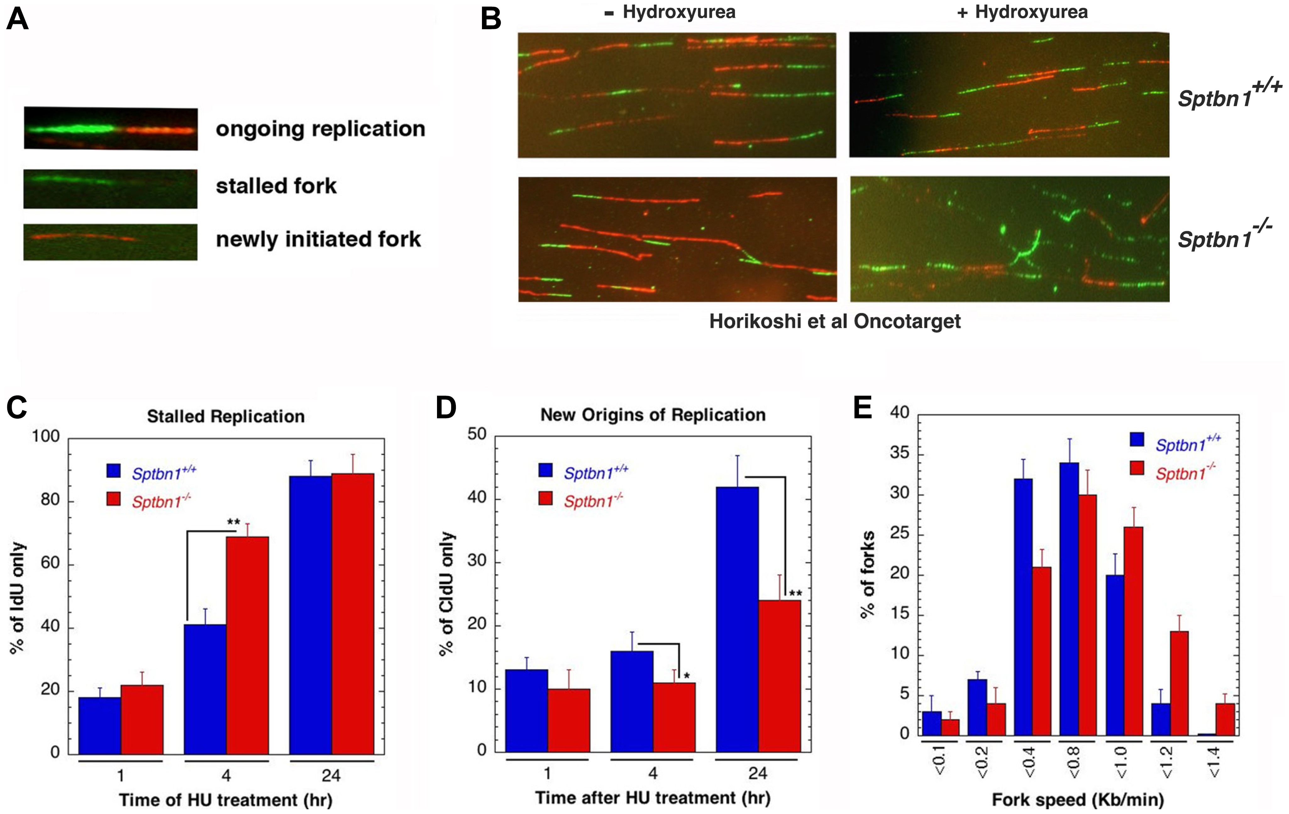
Figure 6: Sptbn1−/− MEFs exhibit defective stalled DNA replication fork resolution and new origins of replication. (A, B) Initiation of new DNA replication forks and reinitiation of stalled DNA replication forks in Sptbn1−/− and Sptbn1+/+ MEFs. cells were pre-labeled with 5-iododeoxyuridine (IdU), treated with hydroxyurea (HU) for the indicated intervals, and then rinsed to remove HU followed by post labeling with 5-chlorodeoxyuridine (CldU) (upper panel) as described previously [66]. The cells were fixed and immunostained with IdU (green) and CldU (red) antibodies. (A) Three major types of labeled DNA tracts for analysis are shown. (B) Sptbn1+/+ and Sptbn1−/− MEFs with and without treatment of hydroxyurea. (C) Percentages of stalled DNA replication forks (IdU only signals) in Sptbn1−/− and Sptbn1+/+ cells after HU treatment. (D) New origins (CldU signals) in Sptbn1−/− and Sptbn1+/+ cells. (E) Percentage of forks with fork speed in Sptbn1−/− and Sptbn1+/+ MEFs. Means ± standard deviations of 3 independent experiments are shown in. *p < 0.05; **p < 0.01, Student t-test.