Corrections:
Correction: IGF-1 and hyperglycaemia-induced FOXA1 and IGFBP-2 affect epithelial to mesenchymal transition in prostate epithelial cells
Metrics: PDF 1470 views | ?
1IGFs and Metabolic Endocrinology Group, Bristol Medical School, Translational Health Sciences, University of Bristol, Southmead Hospital, Bristol, UK
2Faculty of Medicine, Royal College of Medicine Perak, Universiti Kuala Lumpur, Ipoh, MY
3Department of Urology, Bristol Urological Institute, Southmead Hospital, Bristol, UK
4Department of Cellular Pathology, North Bristol NHS Trust, Southmead Hospital, Bristol, UK
5NIHR Biomedical Research Centre, Level 3, University Hospitals Bristol Education Centre, Bristol, UK
6Population Health Sciences, University of Bristol, Bristol, UK
Published: January 26, 2023
Copyright: © 2023 Mansor et al. This is an open access article distributed under the terms of the Creative Commons Attribution License (CC BY 4.0), which permits unrestricted use, distribution, and reproduction in any medium, provided the original author and source are credited.
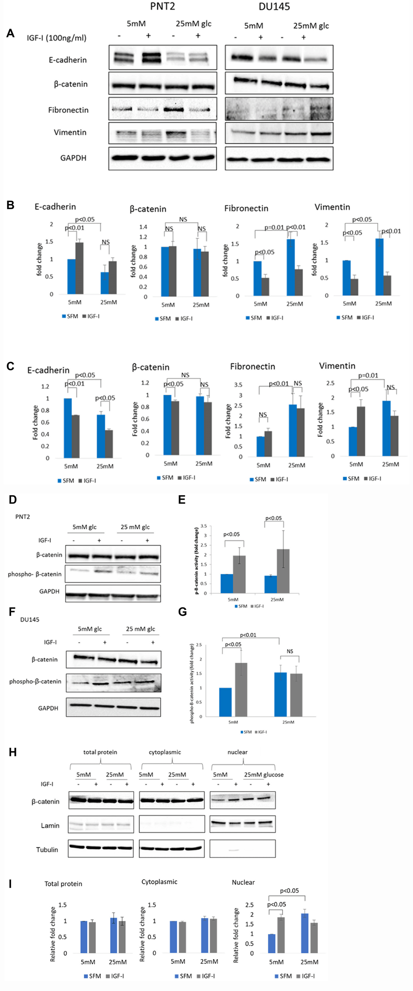
This article has been corrected: In Figure 1, the legend has been amended to show that the β-catenin blots in panel A were re-used in panels D and F as well. The corrected Figure 1 legend is shown below. The authors declare that these corrections do not change the results or conclusions of this paper.
Original article: Oncotarget. 2020; 11:2543–2559. DOI: https://doi.org/10.18632/oncotarget.27650

Figure 1: The effect of IGF-I on EMT markers in prostate epithelial cells in altered glucose condition. (A) Western blot image shows the effect of IGF-I and high glucose on mesenchymal markers in PNT2 and DU145 cells. Cells were dosed with IGF-I 100 ng/ml for 48 hours in normal (5 mM) and high (25 mM) glucose serum free media. Equal amounts of extracted proteins were separated by SDS-PAGE, blotted to a nitrocellulose membrane and probed with primary antibodies against E-cadherin, β-catenin, fibronectin, vimentin and GAPDH. GAPDH was used as a loading control. Optical densities of protein blots for (B) PNT2 and (C) DU145 were quantitated using image J and normalised to GAPDH. Western blots showing regulation of p-β-catenin in (D) PNT2 and (F) DU145 cells when treated with 100 ng/ml IGF-I in normal (5 mM) and high (25 mM) glucose serum free media. The β-catenin blots in (D) and (F) were reused from β-catenin blots in (A). Optical densities of protein blots for (E) PNT2 and (G) DU145 were quantitated using image J and normalised to GAPDH. Ratio of normalised total β-catenin: p- β-catenin were measured and used as an indicator of β-catenin activity. The data expressed as fold changes relative to control represent mean+/− SE of triplicate experiments. (H) Western blot showing cytosolic and nuclear fractions of protein separated form whole cells lysate (total protein) from DU145 cells treated or untreated with 100 ng/ml IGF-I for 48 hours in normal (5 mM) and high (25 mM) glucose serum free media. Lamin A/C and tubulin act as nuclear and cytoplasmic loading controls respectively. Results shown are representative of three independent experiments. Data are represented as mean ± SEM.
 All site content, except where otherwise noted, is licensed under a Creative Commons Attribution 4.0 License.
PII: 28344

