Corrections:
Correction: Hypoxia-inducible factor 1 upregulation of both VEGF and ANGPTL4 is required to promote the angiogenic phenotype in uveal melanoma
Metrics: PDF 1189 views | ?
1 Wilmer Eye Institute, Johns Hopkins School of Medicine, Baltimore, MD, USA
2 The First Affiliated Hospital of Chongqing Medical University, Chongqing, China
3 Department of Pathology, Johns Hopkins University, School of Medicine, Baltimore, MD, USA
4 Departments of Pediatrics, Medicine, Oncology, Radiation Oncology, Biological Chemistry, and Genetic Medicine, Johns Hopkins University School of Medicine, Baltimore, MD, USA
5 Department of Oncology and Diagnostic Sciences, Greenebaum Cancer Center, University of Maryland, Baltimore, MD, USA
* These authors have contributed equally to this work
Published: March 02, 2021
Copyright: © 2021 Hu et al. This is an open access article distributed under the terms of the Creative Commons Attribution License (CC BY 4.0), which permits unrestricted use, distribution, and reproduction in any medium, provided the original author and source are credited.
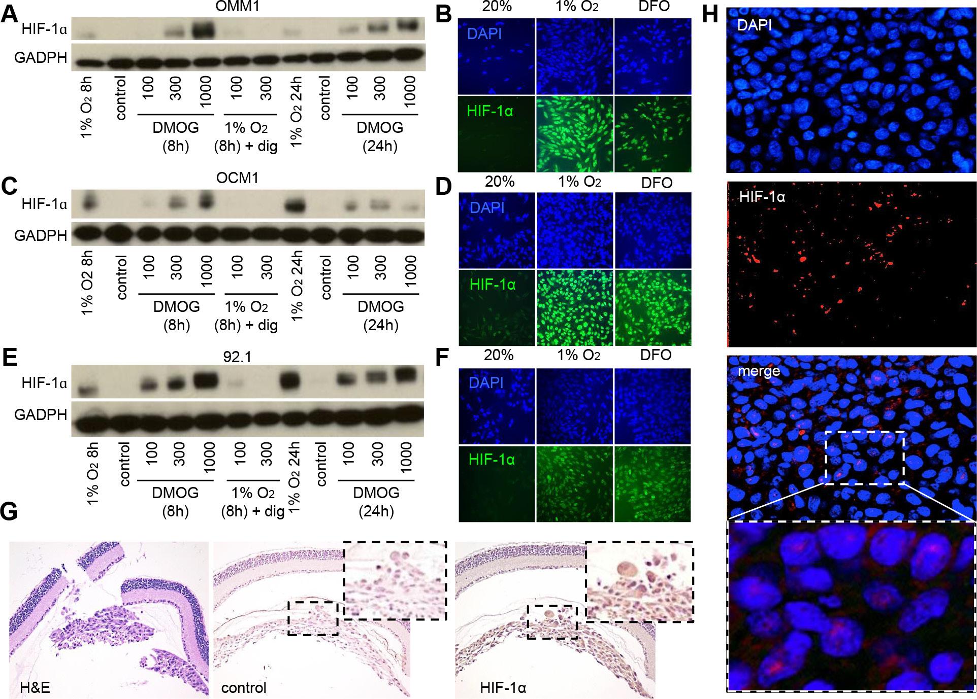
This article has been corrected: In Figure 1, panel D, two of the HIF-1 alpha images were mislabeled. Specifically, the HIF-1 alpha image of OCM1 cells exposed to 1% oxygen was labeled as “DFO” and the HIF-1 alpha image of OCM1 cells exposed to DFO was labeled as “1% oxygen.” The corrected Figure 1 is shown below. The authors declare that these corrections do not change the results or conclusions of this paper.
Original article: Oncotarget. 2016; 7:7816–7828. DOI: https://doi.org/10.18632/oncotarget.6868.

Figure 1: HIF-1α expression is increased in UM cells and in UM patient biopsies. (A, C, E) Immunoblot assays were performed to determine HIF-1α protein levels in UM cell lines (OMM1, OCM1 and 92.1) following exposure to DMOG (300 μM), hypoxia (1% O2) or hypoxia and digoxin (dig; 100-300 nM) for 8 or 24 hours and compared to control conditions (20% O2). (B, D, F) Representative images are shown from immunofluorescence analysis of HIF-1α in UM cell lines following exposure to hypoxia (1% O2 for 8 or 24 hours)or DFO (100 μM for 8 or 24 hours). (G) Representative images are shown from immunohistochemical analysis of HIF-1α expression in tumors formed following intravitreal injection of OCM1 cells into mice. Similar results were observed in 3/3 tumors analyzed. (H) Representative images are shown from immunofluorescence analysis of HIF-1α protein accumulation and nuclear localization in a human UM tumor biopsy. Similar results were observed in 6/6 UM biopsies examined.
 All site content, except where otherwise noted, is licensed under a Creative Commons Attribution 4.0 License.
PII: 27780