Corrections:
Correction: Regulation of E-cadherin localization by microtubule targeting agents: rapid promotion of cortical E-cadherin through p130CAS/Src inhibition by eribulin
Metrics: PDF 1038 views | ?
1 Department of Pharmacology, University of Texas Health Science Center at San Antonio, San Antonio, Texas, USA
2 UT Health Cancer Center, University of Texas Health Science Center at San Antonio, San Antonio, Texas, USA
Published: July 23, 2019
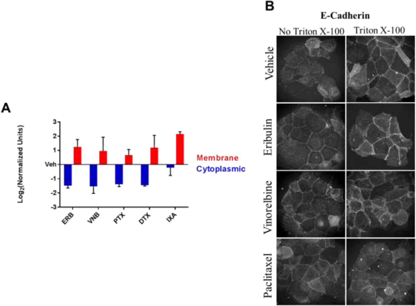
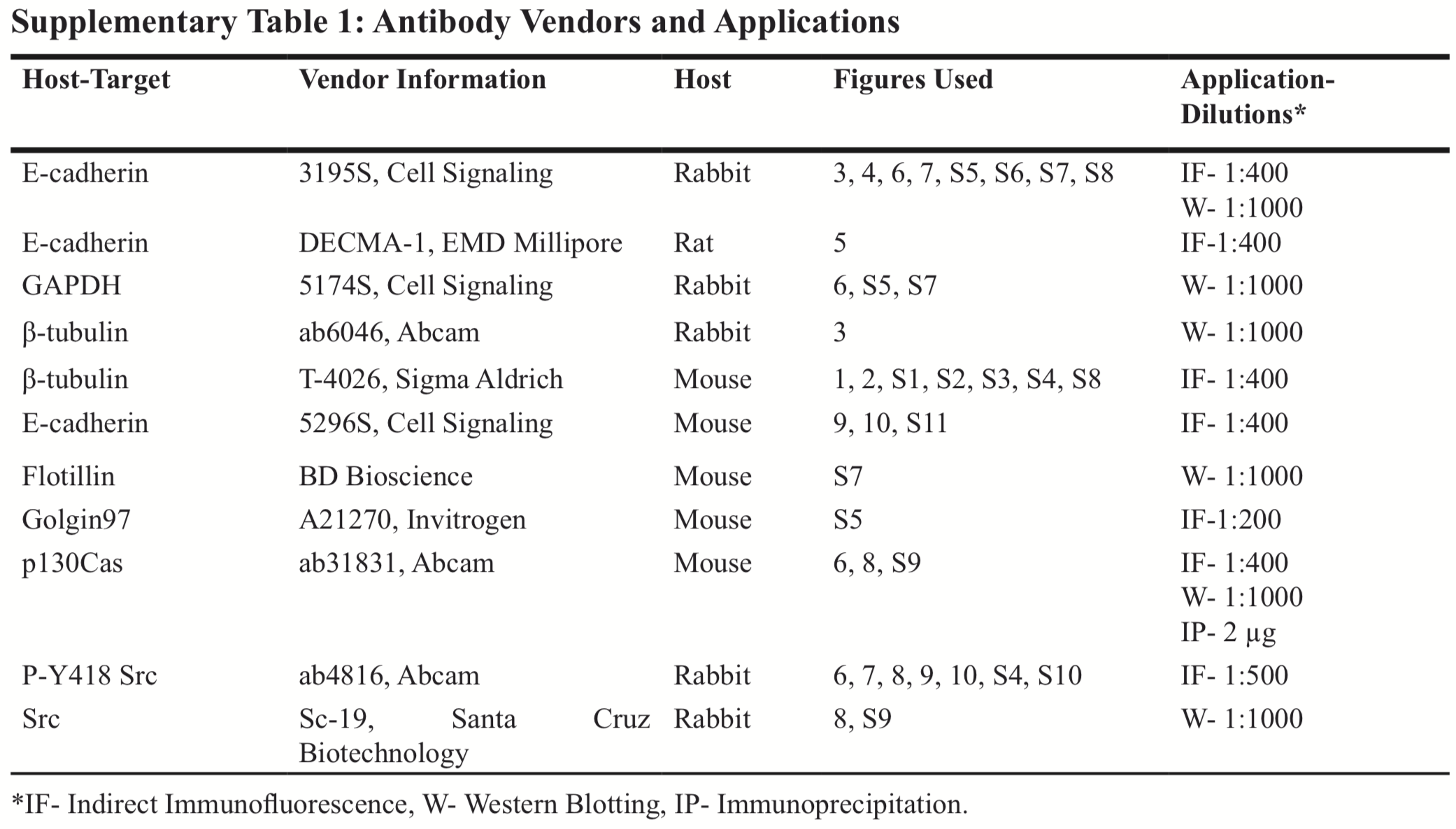
This article has been corrected: The authors recently became aware that the antibody used for Figure 5B was inappropriate to address the ability of E-cadherin to be accessible from the extracellular space in the absence of Triton X permeabilization. The E-cadherin antibody, DECMA-1 from EMD Millipore (Cat. # MABT26) which has the ability to block E-cadherin- associated cell adhesion, is appropriate to address this question. Multiple experiments were conducted with the DECMA-1 antibody in the presence and absence of Triton X permeabilization. New pictures were taken, and a revised Figure 5B using these new images is shown below. A revised Supplementary Table 1 with a list of the antibodies used for each figure is also shown. The authors declare that these corrections do not change the results or conclusions of this paper.
Original article: Oncotarget. 2017; 9:5545–5561. DOI: https://doi.org/10.18632/oncotarget.23798.

Figure 5: The effect of MTAs on the subcellular distribution of E-cadherin. A. Membrane and cytoplasmic-enriched lysates of HCC1937 cells treated for 2 hours with vehicle or MTAs were prepared and analyzed by immunoblotting. Quantification of E-cadherin in the cytoplasmic and membrane-enriched fractions as compared to vehicle. N = 3 ± SEM. B. HCC1937 cells were prepared for indirect immunofluorescence with or without Triton X-100 permeabilization following 4% paraformaldehyde fixation. Arrows indicate E-cadherin ridges between cells in the absence of Triton X-100. Images are composed of non-deconvolved stacks.

 All site content, except where otherwise noted, is licensed under a Creative Commons Attribution 4.0 License.
PII: 27100