


PD-L1 expression in medulloblastoma: an evaluation by subgroup
2018-04-30
The cover for issue 27 of Oncotarget features Figure 5, "MYC overexpression in DAOY (YM21) does not alter PD-L1 expression," from Martin, et al.
This study evaluated the expression of PD‐L1 and markers of immune mediated resistance in human medulloblastoma, the most common malignant pediatric brain tumor.
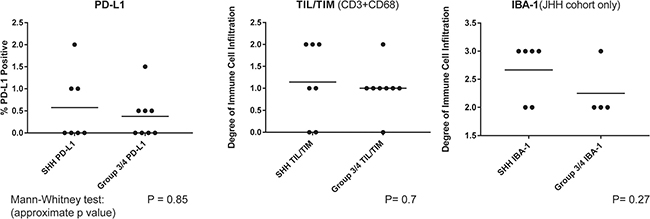
In cell lines, SHH MB, which are low‐MYC expressing, demonstrated both constitutive and inducible expression of PD‐L1 while those in Group 3/4 that expressed high levels of MYC had only inducible expression.
MB expresses low levels of PD‐L1 facilitating immune escape.
"Tumors in the SHH subgroup are characterized by genetic alterations activating this key developmental pathway. WNT subgroup tumors have alterations in the wingless/ ‐catenin developmental pathway."
Most patients are assessed for PD‐L1 expression prior to starting therapy, but the expression of PD‐L1 during the entire course of treatment remains unclear, as does the relationship between changing PD‐L1 expression and therapeutic responses.
In support of PD‐L1 pathway activity in human MB, the researchers demonstrated that MB cell lines robustly up‐regulated PD‐L1 when they simulated an anti‐tumor immune response in vitro by exposing the cell lines to recombinant human IFN−.
In further support of the notion that MB adaptively up‐regulates PD‐L1 as a specific response to immune mediated stimulation is the finding that radiation induced PD‐L1 expression but not to the extent generated by IFN−.
The finding that both IFN− and radiation induced PD‐L1 expression in vitro and the paucity of PD‐L1 expression in vivo in the absence of TIL further emphasizes the concept that immune adjuvants will likely be needed to fully realize the benefit of PD‐1 blockade in cold tumors such as MB.
Expression of MHC II by MB is unusual as this molecule is usually a feature of dendritic cells and other APCs indicating that this tumor may be directly inhibiting anti‐tumor immune responses by masquerading as an inhibitory APC MHC II expression may also indicate a role for the immune checkpoint molecule, lymphocyte activating gene‐3 in MB whose primary ligand is MHC II.
Full text ‐ https://www.oncotarget.com/article/24951/text/
Correspondence to ‐ Allison M. Martin at [email protected]
Copyright © 2026 Rapamycin Press LLC dba Impact Journals
Oncotarget ® is a registered trademark of Rapamycin Press LLC
Impact Journals ® is a registered trademark of Rapamycin Press LLC
RAPAMYCIN PRESS ® is a registered trademark of Rapamycin Press LLC