The cover for issue 66 of Oncotarget features Figure 1, "HD-SCA sample processing and CRC analysis overview," by Gerdtsson, et al.
The research team has established an approach for measuring patient-specific circulating and solid cell concordance by introducing tumor touch preparations to the High-Definition Single Cell Analysis workflow for high-resolution cytomorphometric characterization of metastatic colorectal cancer.
Combined with the association of circulating cells with tumor burden and necrosis of hepatic lesions, their overall findings demonstrate that liquid biopsy cells can be informative biomarkers in the mCRC setting.
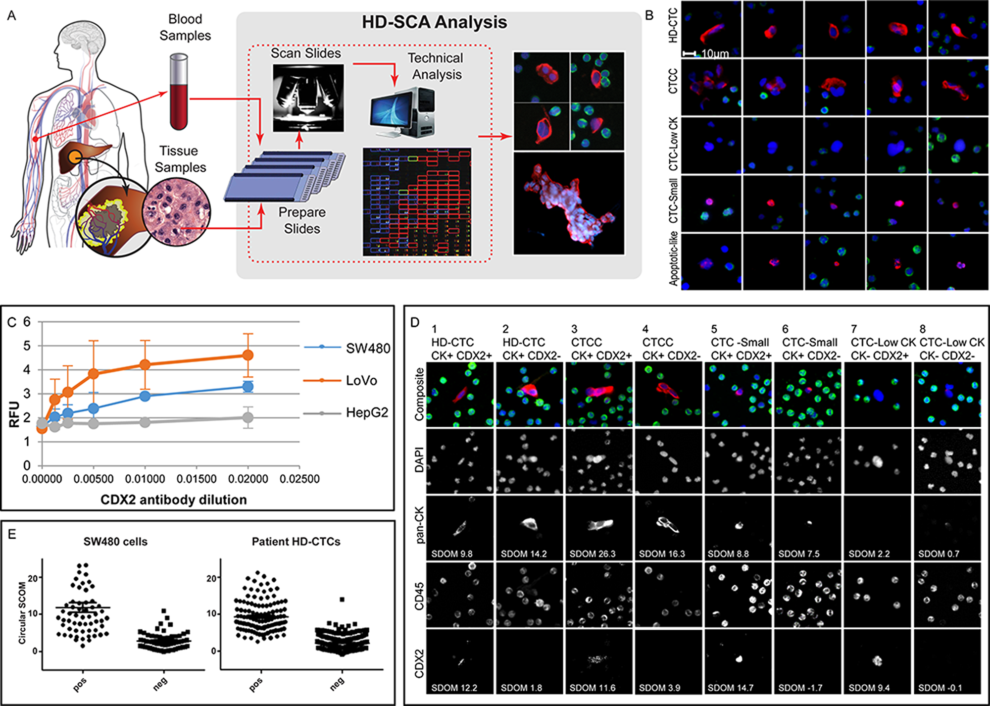
Figure 1: HD-SCA sample processing and CRC analysis overview. (A) Peripheral blood (liquid biopsies) collected prior to surgery and tumor touch preps (solid biopsies) collected from each piece of resected liver (or colon) tumor were processed, stained, and imaged in parallel. Candidate tumor cells were re-imaged at higher resolution and segmented for collection of single cell imaging data. (B) Cells of different categories as classified in the HD-SCA report system identified in mCRC patients. Each row of composite images represents one cell category (DAPI, blue; pan-CK, red; CD45, green). HD-CTCs and CTCCs were included in the analysis. Other cell categories classified in the HD-SCA report system are CTC-Low CK (CK-/low, CD45- morphologically distinct and larger than surrounding WBCs), CTC-Small (CK+, CD45- with similar size as surrounding WBCs), Apoptotic-like (CK+, CD45- cells with irregular or disrupted nucleus and/or cytoplasm) as well as False Positives and WBCs (not shown). (C) A CDX2 monoclonal antibody was selected after an initial antibody screening using plated positive (SW480 and LoVo) and negative (HepG2) control cell lines and titrated in 3 independent experiments (RFU=Relative Fluorescence Units). (D) SW480 cells were spiked in whole blood and processed according to the HD-SCA protocol. The CDX2 SDOM of tumor cells relative to surrounding WBCs was similarly distributed for CDX2+ and CDX2- cells in spiked SW480 cells and patient HD-CTCs from the first HD-CTC positive patient (CRC004). (E) Examples of cell subgroups as defined by HD-SCA cell category and CK and CDX2 expression, identified from a single sample from patient CRC112. Images were taken at 40X magnification, except for cell 7 where images were taken directly from the 10X scanning report due to lack of 40X imagery. Each column depicts the target cell(s) with a composite image (DAPI, blue; pan-CK, red; CD45, green) on top and the individual channels below.
Patient-specific level of concordance can readily be measured to establish the utility of circulating cells as biomarkers and define biosignatures for liquid biopsy assays.
Dr. Peter Kuhn from the USC Michelson Center for Convergent Bioscience, at the University of Southern California, Los Angeles, CA, USA said, "Colorectal cancer (CRC) is the third most common cancer globally."
To faithfully capture cell heterogeneity, personalized oncology is furthermore moving towards single cell profiling for more informed clinical decision making, as opposed to homogenized cell populations in conventional bulk biopsy analysis.
High-Definition Single Cell Analysis is a workflow for detailed characterization of circulating rare cells by high resolution imagery of individual cells.
By screening all nucleated cells within a sample, it is a "no cell left behind" approach enabling both increased sensitivity compared to most other CTC assays, as well as inherent morphometric profiling of all candidate tumor cells.
With an earlier version of the workflow, a case study of CTCs and primary cells from lung cancer concluded that a subset of CTCs displayed similar cytomorphology to a subset of primary tumor cells, even though the solid biopsy had been collected years prior to the CTCs.
Based on the current HD-SCA workflow, which includes a more detailed digital analysis of candidate cells and allows for incorporating solid biopsy single cell analysis, the authors set out to establish an approach for quantitative correlation analysis of liquid and solid biopsy cells based on high-content morphometric analysis on the single-cell level.
The Kuhn Research Team concluded that while the study was limited by the current lack of genomic and/or multiplex proteomic single-cell data, the morphometric characterization of >1000 single cells supports that liquid biopsy cells are related to solid tumor cells in mCRC, and that subgroups of cells with characteristic morphology are present across patients and the liquid and solid cell compartments.
Analysis of tumor touch preparations in parallel with the conventional liquid biopsy cell profiling within the HD-SCA platform can be readily introduced to the workflow to assess the level of concordance within a patient, and to determine the applicability of liquid biopsies for tumor characterization for each individual case.
Full text - https://doi.org/10.18632/oncotarget.27271
Correspondence to - Peter Kuhn - [email protected]
Keywords - single cell analysis, liquid biopsy, metastatic colorectal cancer, circulating tumor cells, cytomorphology