Corrections:
Correction: Inhibition of ATM kinase upregulates levels of cell death induced by cannabidiol and γ-irradiation in human glioblastoma cells
Metrics: PDF 1177 views | ?
1 Center for Radiological Research, Department of Radiation Oncology, Vagelos College of Physicians and Surgeons, Columbia University, New York, NY 10032, USA
Published: December 10, 2019
This article has been corrected: Due to errors during figure assembly, the image used in Figure 3C is incorrect. In addition, the image for Figure 3A contains accidental duplication of FACS panels. The proper Figure 3 is shown below. The authors declare that these corrections do not change the results or conclusions of this paper.
Original article: Oncotarget. 2019; 10:825–846. DOI: https://doi.org/10.18632/oncotarget.26582.
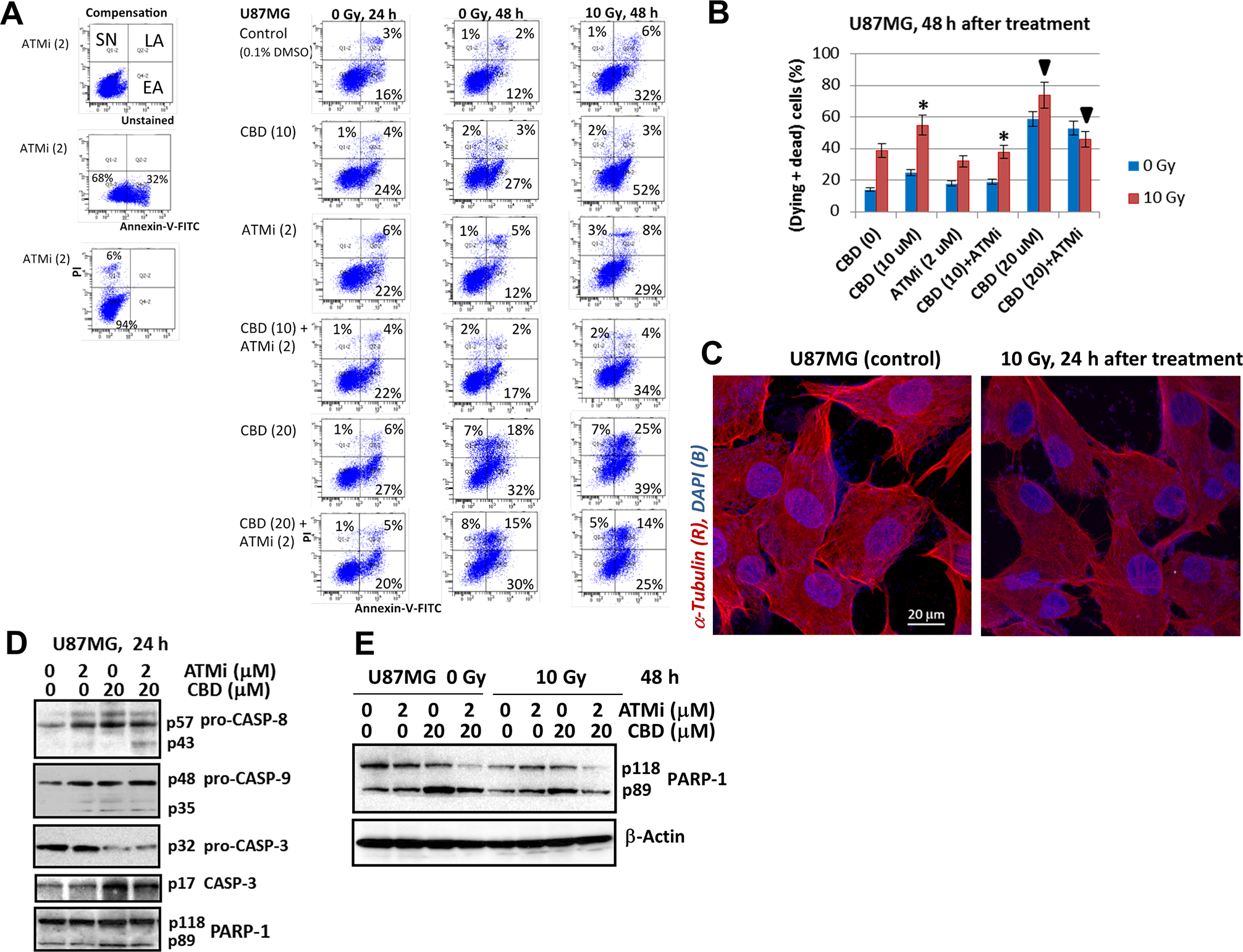
Figure 3: The apoptotic commitment of U87MG after treatment with CBD (10-20 μM), ATMi (2 μM) and γ-irradiation (10 Gy), alone or in combinations. (A and B) Annexin-V-FITC and PI staining for determination of early apoptotic (EA), late apoptotic (LA) and secondary-necrotic (SN) GBM cells after indicated treatment was followed by the flow cytometry. Typical experiment (A) and pooled results of four independent experiments (B) using U87MG cells 24-48 h after indicated treatments are shown. Percentage of (dying + dead cells) included early apoptotic (EA), late apoptotic (LA) and secondary necrotic cells (SN). Error bars represent means ± S.D. (p < 0.05, Student’s t-test). The stars and the arrows indicate significant differences between indicated cells after specified treatment. (C) The images of control and irradiated U87MG after immunostaining with α-Tubulin and DAPI followed by confocal microscopy are shown. (D and E) Western blot analysis of apoptotic marker proteins 24 h and 48 h after indicated treatments of U87MG cells.
 All site content, except where otherwise noted, is licensed under a Creative Commons Attribution 4.0 License.
PII: 27352