Corrections:
Correction: Carcinoma-risk variant of EBNA1 deregulates Epstein-Barr Virus episomal latency
Metrics: PDF 1246 views | ?
1 The Wistar Institute, Philadelphia, PA USA
2 Deutsches Krebsforschungszentrum, Heidelberg, Germany
3 Department of Pediatrics and Adolescent Medicine, The University of Hong Kong, Hong Kong
Published: July 09, 2019
This article has been corrected: Due to errors in image selection, Figure 5A shows an inadvertent duplicate of Donor 2 for M81. Figure 7D shows the Actin control from Donor 2 that was inadvertently duplicated from the Donor 2 Actin control shown in Figure 4 (which is the same extract and donor sample). Figure 4 is correct and unchanged. The corrected Figure 5 and Figure 7 are shown below. The authors declare that these corrections do not change the results or conclusions of this paper.
Original article: Oncotarget. 2017; 8:7248-7264. DOI: https://doi.org/10.18632/oncotarget.14540.
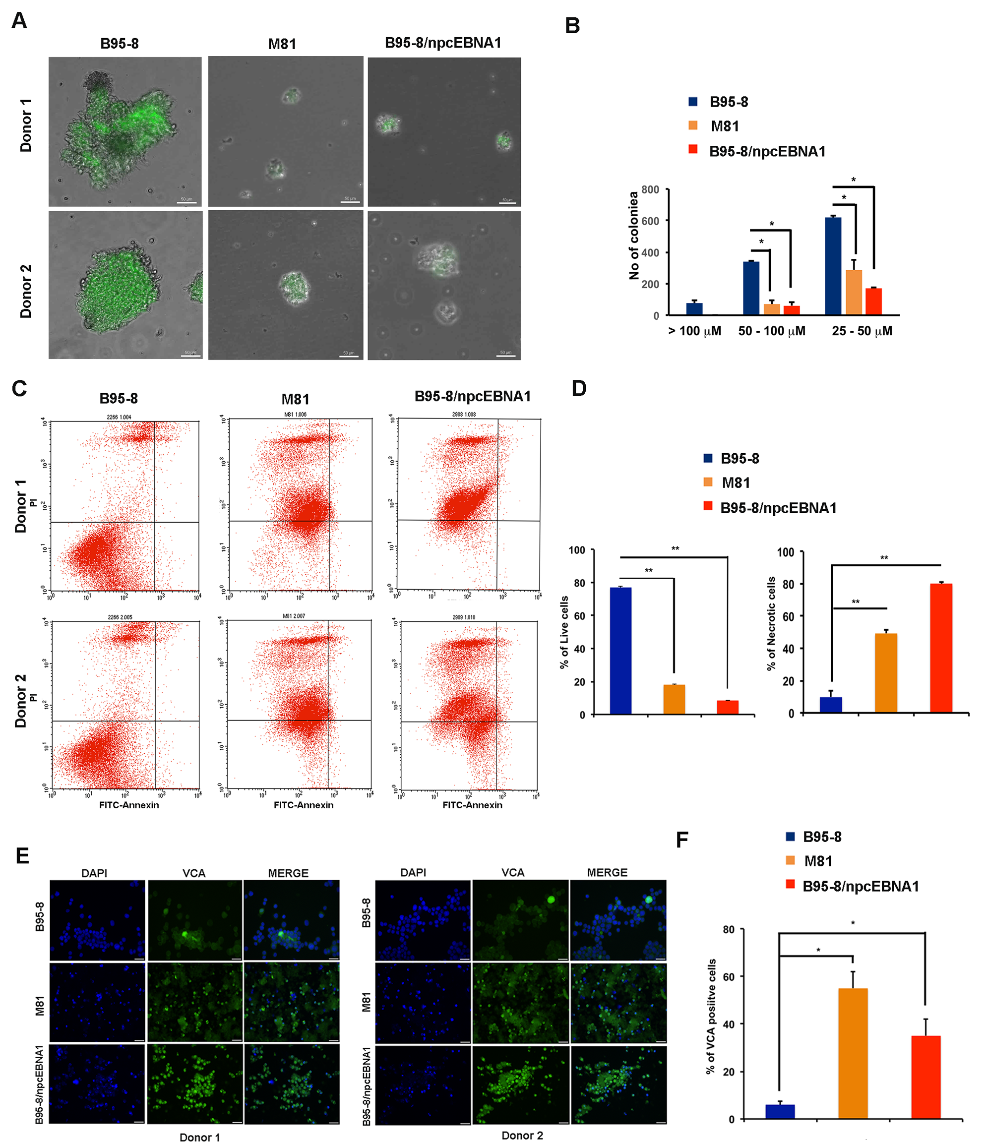
Figure 5: Defective B-cell blast formation by B95-8/npcEBNA1. Bacmid-derived virus for B95-8, M81, or B95-8/npcEBNA1 were assayed at 2 weeks post-infection of primary B-lymphocytes. A. GFP positive B-cell blasts for two independent donors were analyzed by high-throughput microscopy. B. The number of colonies imaged by microscopy with diameters of > 100, 50-100, or 25-100μM were quantified by Image J (panel B). C. B-cell blasts at 4 weeks post-infection were analyzed by FACS for apoptosis using propidium idodide (PI) (x-axis) and annexin V (y-axis). D. The percentage of live and necrotic cells assayed by FACS were quantified for two independent donors and three independent biological replicates.
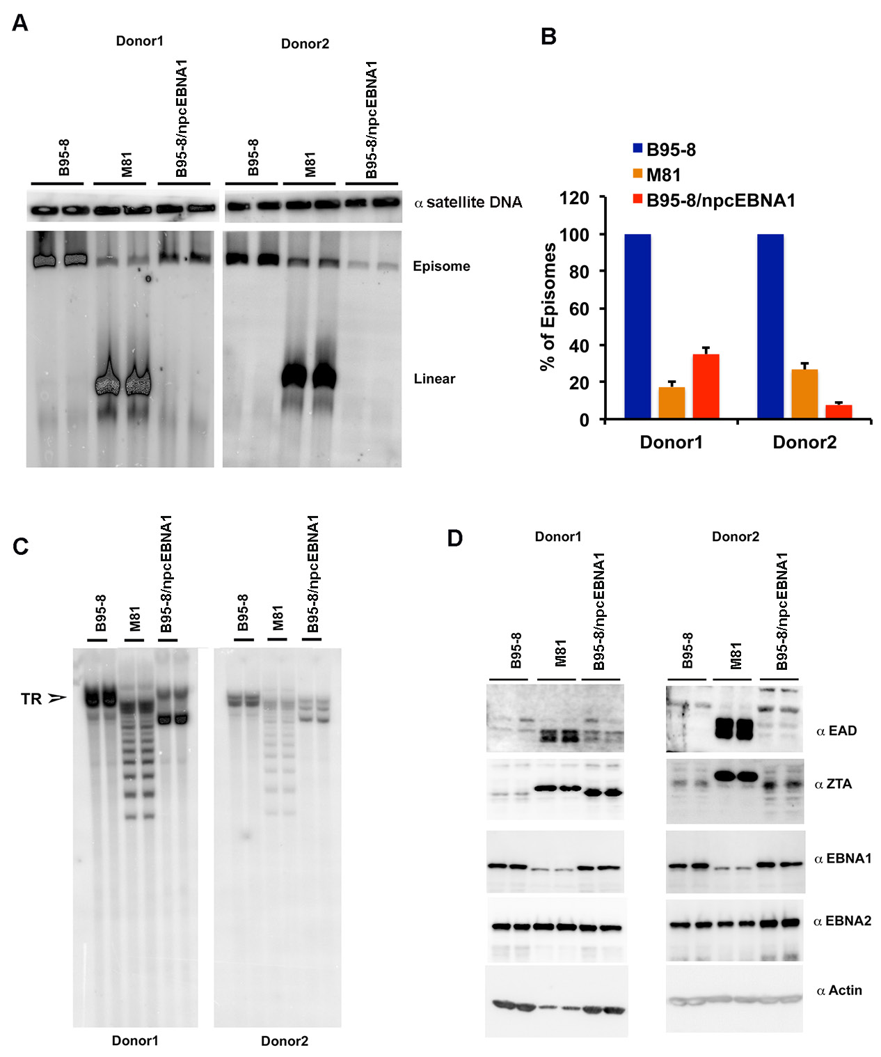
Figure 7: Low episome copy number and terminal repeat instability in LCLs with B95-8/npcEBNA1. A. PFGE analysis of LCLs generated with recombinant B95-8, M81, or B95-8/npcEBNA1 virus. Samples are run as technical replicates for two independent donor generated LCLs. Cellular α-satellite DNA is shown as loading control above each lane. B. Quantitation of EBV episomes relative to α-satellite DNA for PFGE shown in panel A. C. Southern blot analysis of EBV terminal repeats after digestion with BamHI for B95-8, M81, or B95-8/npcEBNA1 generated LCLs. D. Western blot for EAD, ZTA, EBNA1, EBNA2, and Actin for B95-8, M81, or B95-8/npcEBNA1 generated LCLs.
 All site content, except where otherwise noted, is licensed under a Creative Commons Attribution 4.0 License.
PII: 27073